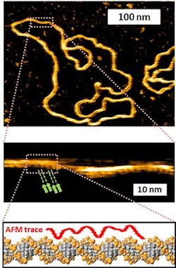
Atomic force microscopy captures first images of DNA's double helix in water
Researchers at the London Centre for Nanotechnology, led by Bart Hoogenboom, Ph.D., have used atomic force microscopy (AFM) to capture the first images of DNA's double helix in water.
Hoogenboom's main research interest is where nanotechnology tools could be used to study and manipulate single biomolecules. He has identified AFM as the only instrument that provides ~1 nm spatial resolution on large biomolecules that are still functional and may be studied in the natural environment, such as in aqueous salt solution. To that end, AFM can visualize biological processes while they happen, and at the scale of single molecules. It also enables him to study electrostatic charge and nanomechanical rigidity.
Hoogenboom notes that the AFMs they use (JPK Instruments' NanoWizard AFMs) are compatible with inverted optical microscopes, which enable larger-scale images of what they are viewing at the nanoscale. For experiments at higher spatial and time resolution, they also use home-built microscopes, he says. While using these microscopes, JPK has supplied them with its Vortis AFM controller.
Most current microscopes do not achieve any higher resolution on DNA than was achieved with the first AFM experiments in the early 1990s, says Hoogenboom.
The work has been published in Nano Letters; for more information, please visit http://pubs.acs.org/doi/abs/10.1021/nl301857p.
-----
Follow us on Twitter, 'like' us on Facebook, and join our group on LinkedIn
Laser Focus World has gone mobile: Get all of the mobile-friendly options here.
Subscribe now to BioOptics World magazine; it's free!

