YONG-LE PAN and RICHARD K. CHANG
The photomultiplier tube (PMT) is well-known as a single-photon-sensitive detector that measures the intensity as a function of time, I(t), well into the gigahertz range. The PMT cannot, however, measure the spatial distribution of the intensity. Nevertheless, the PMT is one of the detectors most frequently used in laboratory experiments and optical instrumentation. By contrast, a charge-coupled-device (CCD) detector—because of its numerous pixels—can measure the spatial variation of the intensity I(R), but cannot measure frame changes at rates greater than 100 Hz.
Standard PMT characteristics
A standard PMT consists of a large-area photocathode that converts photons to photoelectrons via the photoelectric effect. No matter where the incoming photons strike the photocathode, electrostatic fields direct the resulting photoelectrons toward a single electron-multiplier unit. Electron-number gain (as much as 106 to 108) usually is achieved with 9 to 12 dynodes that produce secondary electrons. All the secondary electrons, which result from a single photoelectron, arrive at the anode as a packet with a temporal width (typically 1 ns) that is ultimately limited by the spread in the arrival time of the secondary electrons at the anode. Such a spread is caused by the slightly different trajectories the second electronics take to reach the anode from the first dynode. The anode is usually terminated by 50 Ω in fast-response PMTs to minimize the distortion of the output photocurrent pulse shape, by matching with the impedance of the coaxial cable. Hence, the output-voltage pulse is 50 Ω times the anode-current pulse.1
The PMT is said to be single-photon-sensitive or quantum-limited if the current- or voltage-pulse height associated with one photoelectron from the photocathode is greater than the pulses from photo- or thermionic-electron emission from the dynodes. There is no way to distinguish between the current and voltage pulses, however, due to the emissions from the photocathode. Thermionic-electron emission is commonly referred to as "dark noise" or "dark current."
Multi-anode PMT characteristics
The multiple-anode PMT is equivalent to incorporating multiple PMTs in a single housing. An output pin protrudes from the base of the housing for each of the 32 anodes, which means that each anode is externally accessible. The uniqueness of the multiple-anode PMT is in the electron-multiplication section—it is distributed throughout the housing. The objective of this section is to achieve nine stages of electron multiplication (a gain of nearly 106) while preserving the spatial integrity of a photoelectron that is emitted from a particular location (x, y) on the photocathode. This ability allows for a large variety of multiple-anode PMTs.
The conventional PMT is a zero-dimensional detector, in which the spatial information of the intensity along the x- and y- axes is lost and summed by one large anode that intercepts all the secondary electrons. The 32-anode PMT provides one-dimensional (1-D) information—the spatial information of the intensity along the y-axis is lost and summed by the narrow-stripe anode. A 64-anode PMT with an 8 x 8 square array of anodes and a square-shaped photocathode (1.81 x 1.81 cm) also exists, providing coarse two-dimensional intensity information.
In the 32-anode PMT gain can vary by as much as 10% among the anodes. Such gain variations are caused by nonuniformity of the photocathode sensitivity and/or of the distributed-dynode gain. As much as 1% crosstalk between the adjacent anodes is unavoidable, and is caused by leakage of electrons from one stripe (x) within the distributed electron-multiplication unit to its neighboring stripes (x ± Δx). For the nearest neighboring anodes, crosstalk is much less—as little as 0.01%.
Three experimental configurations
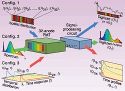
The availability of a 1-D 32-anode PMT means that various types of experimental configurations can be set up using a single device instead of many conventional PMTs. The first configuration demonstrates the uniqueness of the multiple-anode PMT when the intensity at many widely separated locations needs to be measured simultaneously—in an elastic scattering experiment of microparticles, for example, the scattered intensity at many different angles (q and F) must be measured simultaneously. In this example, individual fibers carry the intensity information from different locations. Placing these fibers side-by-side along the x-axis of the PMT and imaging them onto the photocathode allows the different anodes to monitor the intensity at various locations.
The second configuration is applicable when the spectral lineshape of a single peak, or the intensity of several peaks, must be recorded simultaneously from a flowing aerosol. The photocathode of the 32-anode PMT is placed at the focal plane of a spectrograph, with the groves/mm of its grating optimized for linewidth determination, as well as for the overall spectral coverage considerations.
When the temporal response at several different wavelengths must be measured simultaneously, the third configuration becomes valuable. Again, the multiple-anode PMT is placed at the focal plane of the spectrograph, allowing temporal responses of several anodes to be recorded with a multichannel oscilloscope.
Fluorescence sensing
We have used the 32-anode PMT to determine the fluorescence spectrum of individual bio-aerosols flowing rapidly (1 to 10 m/s) through the focal volume of an ultraviolet (UV) laser beam.1 The entire fluorescence spectrum from the aerosol, excited at 266 nm by the UV laser, can consist of several peaks and can extend from 200 to 700 nm. We wanted to detect the entire spectrum and store it in a buffer memory for each 70-ns laser pulse. The laser was triggered to fire only when a particle randomly flowed through the sample volume.
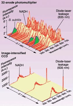
In our experiment, which highlights the speed advantage of the 32-anode PMT, the entire spectral data from the 32 wavelength bands could be stored into buffer memory as fast as 1.4 kHz, which compares with the 25-Hz frame rate of an image-intensified-CCD (ICCD) detector. Consecutive single-shot fluorescence spectra of a mixture of two types of aerosolized particles (~5 µm in diameter)—Bacillus subtilis bacteria and nicotinamide adenine dinucleitide (NADH) aggregates—were measured (see Fig. 2). Because of the difference in the number density of the two types of particles, only three B. subtilis bacteria spectra (peaked at 330 nm) were detected while dominated by NADH aerosols (97 spectra, peaked at 450 nm). The sharp peak at 635 nm is the elastic scattering from the semiconductor-diode laser that is used to determine the random arrival of aerosols at the trigger volume, which is just upstream from the sample volume.Vtech Engineering Corp. (Andover, MA) designed and constructed the data-acquisition and processing system (PhotoniQ P7260-1) for the 32-anode PMT. System-control software was written in C++.2 We systematically characterized performance of the 32-anode PMT at room temperature and determined its data frame rate (1.4 kHz) and dynamic range (better than 103). Its sensitivity was comparable to an ICCD detector in terms of being able to detect and read single-photon-electron events.
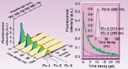
To illustrate the temporal behavior of the PMT at several wavelengths, we directly measured the voltage pulse at several anode pins, which were selected based on their correspondence with the wavelengths of interest (see Fig. 3). In the spectra, the sharp peak at 266 nm is the leakage of elastic scattering from the excitation laser. The peaks around 650 nm are the second-order grating response of the fluorescence around 320 nm. The lithium dioxide (LiO2) fluorescence spectra consist of two broad bands (maximums at 320 and 370 nm), and their relative intensity varies with delay time. The two fluorescence bands suggest that the emissions result from two different electronic-energy levels with different lifetimes. The stronger band at 320 nm has a much shorter lifetime than the weaker band at 370 nm.To characterize this spectral change as a function of time delay, we directly measured the fluorescence decay time of the 320- and the 370-nm bands as well as the pulse duration of the 266-nm pump pulse (see Fig. 3, right). All three time-responses were measured simultaneously by the 32-anode PMT. Temporal behavior at any other wavelength could be easily measured simply by connecting to a different anode pin, without the need to turn the grating or move several conventional PMTs to different locations along the focal plane of the spectrograph. The temporal profile indicates that the laser is a 12-ns broad pulse. The fluorescence at 310 nm has a short lifetime of less than 5 ns and closely follows the laser pulse. The fluorescence at 390 nm has a long lifetime of about 6 µs and covers a wavelength range from 300 to 450 nm.
The difference in temporal response of different anodes demonstrates that the multiple-anode PMT combines the advantages of a CCD (spatial resolution) and the conventional PMT (fast response time). The PMT has fewer pixels, by orders of magnitude, than a standard CCD detector. The multiple-anode PMT, however, has time-response in the gigahertz range, while the CCD detector has a frame rate in the 100-Hz range. It is this dual capability (spatial and temporal) of the multiple-anode PMT that makes it such a desirable and unique detector.
ACKNOWLEDGMENT
The authors wish to acknowledge informative discussions with Dr. Ken Kaufmann and Dr. Norman Schiller of Hamamatsu Corp. (Bridgewater, NJ). The expertise of and collaboration with Prof. Jean-Pierre Wolf of Universite Claude Bernard Lyon, France; Vtech Engineering Corp. (Patrick Cobler and Scott Rhodes); and Dr. Ronald G. Pinnick of the US Army Research Laboratory have been most helpful. The partial financial support of the US Army Research Office (DAAG55-97-1-0349) and US Army Research Laboratory (ARL contract DAAL01-98-C0056) is gratefully acknowledged.
REFERENCES
- Hamamatsu Photonics K. K editorial committee, Photomultiplier Tube Principle to Application (Japan, 1994).
- Y. L. Pan et al., Rev. Sci. Instrum. 72, 1831 (2001).
Yong-Le Pan is a research physicist with the Physical Sciences Lab, New Mexico State University, Las Cruces, NM 88003; e-mail: [email protected]. Richard K. Chang is the Henry Ford II Professor in the Department of Applied Physics, Yale University, New Haven, CT 06520; e-mail: [email protected].


