FIBER-BASED SENSORS: Surface-enhanced Raman sensors improve detection of dangerous agents
CLAIRE GU and CHAO SHI
The demand for sensors capable of detecting chemical and biological agents is greater than ever, arising from applications that include medical, environmental, food safety, military, and security. Currently, most detection or sensing techniques tend to be bulky, expensive, relatively inaccurate, or unable to provide real-time data. Clearly, alternative sensing technologies are urgently needed.
Surface-enhanced Raman scattering (SERS), first discovered in the 1970s, provides a promising technology for highly sensitive, molecule-specific detection (see www.laserfocusworld.com/articles/274732).1, 2 Raman scattering carries the information of molecular vibrational energy levels, and therefore, it reveals the intrinsic signature of the molecule. However, the small cross section of Raman scattering limits its sensitivity in practical applications. In SERS, a molecule is adsorbed to a noble-metal surface with nanoscale features, in either films or colloidal suspensions. When the surface-plasmon mode of the metal is excited with a laser, it causes a local field enhancement near the surface, which in turn enhances Raman scattering of molecules adsorbed or in close proximity to the surface. The SERS enhancement factor varies with the substrate nanostructure but can range from 103 to 1015.
Optical fibers have long been used in sensors because of their ability to transmit light in a flexible and compact fashion. In recent years, research has turned to the use of SERS and optical fiber for their potential in chemical, biological, and environmental detection. The combination of SERS and optical fiber offers the advantages of the molecular specificity of Raman scattering, the huge enhancement factor of SERS, and the flexibility of optical fibers. In addition, the confinement of the optical light inside the core of a fiber has a unique advantage in nonlinear processes such as Raman scattering, which require maximization of interaction length and optical intensity.
Three kinds of fiber SERS probes are of interest for sensing. The first is a simple multimode fiber probe, featuring a tip coated with a layer of nanoparticles that serves to enhance the electromagnetic field around the probe. The second is a hollow-core photonic-crystal fiber (HCPCF), the core of which is filled with a mixture of nanoparticle solution and the sample to increase the interaction volume for SERS. The third is a combination of the features in the above two–a liquid-core photonic-crystal fiber with an inner wall coated with a layer of nanoparticles.
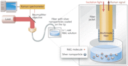
The simple tip-coated multimode fiber (TCMMF) probe is also called a double-substrate SERS probe (see Fig. 1), in which substrate in this sense is nanoparticles around which SERS occurs.3 The approach is to coat the tip of a multimode fiber (MMF) with a SERS substrate solution, such as silver nanoparticles (SNPs). A 633 nm diode-laser source at the SERS excitation wavelength is focused into the fiber and absorbed by the silver nanoparticles on the fiber tip, forming a strong field around the tip. Target analyte molecules such as rhodamine 6G (R6G) are combined in another substrate solution with different SNPs. When the tip of the probe is dipped into the target solution, the solution interacts and binds to the SNPs on the fiber tip. Statistically, some of the molecules are sandwiched in the junction between the two SNP substrates, where the electromagnetic field is further enhanced, leading to stronger SERS signals. The SERS signal from the sample propagates back through the multimode fiber toward the source, where the Raman spectrometer collects it.
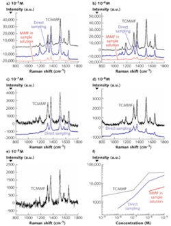
The results using R6G as a testing molecule at varied concentrations are different for the three fiber SERS probes (see Fig. 2). The various testing schemes include: direct detection of the SERS signal in the sample solution; detection with an uncoated MMF probe dipped in the sample solution; detection with the TCMMF probe dipped in an aqueous R6G solution; and detection with the TCMMF probe dipped in the sample solution.The lowest concentration detectable using the tip-coated multimode fiber probe dipped in the aqueous R6G solution was only 10-3 M to 10-4 M, which was much higher than the other three methods. Based on quantitative comparison of the SERS results, the lowest detectable concentrations using the MMF probe, direct-solution detection, and the TCMMF probe, were 10-6 M, 10-8 M, and 10-9 M, respectively.
For the same concentration of R6G, the signal intensity from the TCMMF probe was consistently much higher than that from the MMF probe or direct solution detection, as well as the simple sum of the signals from MMF plus the direct solution detection. This indicates stronger SERS activity with the TCMMF due most likely to stronger electromagnetic enhancement as a result of the unique “sandwich” structure. Such sandwich structures formed by SNPs on the fiber probe and in solution are likely to exhibit stronger SERS due to stronger electromagnetic enhancement compared to each substrate alone since some of the R6G analyte molecules are at junctions of SNPs.
The LCPCF probe
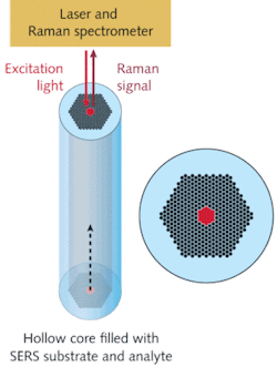
An alternative probe that can also achieve higher sensitivity than direct detection is the liquid-core photonic-crystal-fiber (LCPCF) probe, in which the cladding holes of a hollow-core photonic-crystal fiber are sealed at one end and the central hole is open and filled with liquid samples (see Fig. 3).4 As both the light and the sample are confined in the fiber core, and the photonic bandgap is still preserved, the SERS sensitivity is significantly improved. The newly developed LCPCF sensor was tested in the detection of R6G, human insulin, and tryptophan with good sensitivity due to the enhanced interaction volume.Fiber SERS probes can offer not only flexibility and compactness but also higher sensitivity than that obtained by directly focusing a laser beam into the sample. The mechanism for the increased sensitivity is the use of more SERS particles in the active region, or to further enhance the electromagnetic field around the probe area, or both. Examples of various fiber SERS probes have been demonstrated. The LCPCF confines the excitation light and the liquid sample inside its microcavity, leading to an increased SERS interaction volume. The TCMMF probe takes advantage of the additional electromagnetic field enhancement between the two SERS substrates.
Another benefit of the double-substrate fiber SERS probe is its simple modification of regular multimode fibers, leading to its insensitivity to the excitation wavelength and ease of a practical method for fabrication. A combination of these two approaches results in the inner-wall-coated LCPCF probe, which has even higher sensitivities; therefore, it provides an extremely sensitive SERS sensor. With the integration of fiber SERS probes into a compact sensor system, surface-enhanced Raman sensors are promising for use in highly sensitive, molecular-specific, and low-cost chemical and biological detection applications.
Acknowledgments
The authors would like to thank Yi Zhang, Chao Lu, and He Yan of the Department of Electrical Engineering, and Debraj Ghosh, Leo Seballos, Shaowei Chen, and Jin Z. Zhang with the Department of Chemistry, UC Santa Cruz, for their assistance.
References
- M. G. Albrecht and J. A. Creighton, J. Am. Chem. Soc. 99 (15), p. 5215 (1977).
- L. Jeanmaire and R.P. Vanduyne, J. Electroan. Chem. 84 (1), p. 1 (1977).
- C. Shi et al., Appl. Phys. Lett. 92, p. 103107 (2008).
- Y. Zhang et al., Appl. Phys. Lett. 90, p. 193504 (2007).
- C. Shi et al., to appear in Appl. Phys. Lett. 93, p. 153101 (2008)
Claire Gu is a professor and Chao Shi is a doctoral student in the Department of Electrical Engineering, 253B Baskin Engineering Building, University of California, Santa Cruz, CA 95064; e-mail: claire@ee.ucsc.edu; www.soe.ucsc.edu.


