BIOMEDICAL IMAGING: Holographic live-tissue imaging uses photorefractive polymer

Collection of three-dimensional (3-D) data from confocal scanning microscopy and optical coherence tomography (OCT) is lengthy due to the sequential acquisition of image pixels required for these methods; for this reason, researchers at the University of Cologne (Cologne, Germany) and Purdue University (West Lafayette, IN) are pursuing a variation of holographic optical coherence imaging (HOCI) as a faster depth-resolved imaging technique.1 Because the whole image is formed in one step, lengthy point scanning is avoided. However, another major obstacle for any tissue-imaging system is light scattering and its impact on image resolution. To combat this concern, the research team implemented coherence-gated holography as a means to separate unscattered or image-bearing “ballistic” light from scattered incoherent light.
Previous methods to implement coherence-gated holography for depth-selective optical sampling of biological tissues (and even video) were made possible due to dramatic improvements in the computational processing power of digital holography (DH), in which a holographic interference pattern is digitally recorded by a charge-coupled-device (CCD) camera. However, DH still requires numerical image reconstruction or post-data processing with computed-tomography techniques that can introduce mathematical artifacts. But by replacing DH with highly sensitive photorefractive (PR) holography, image reconstruction becomes purely optical and post-data processing is not required.
Photorefractive holography
An alternative to bulk-crystal or semiconductor-quantum-well-based PR materials is PR organic polymers, which are easier to fabricate, offer improved spatial resolution, and have very high diffraction efficiencies—up to 100% compared to 4% for quantum-well-based semiconductor PRs. Recognizing that low material sensitivity of polymer-based PRs has limited their use, however, the research team developed a PR polymer composite with high near-IR sensitivity and uses it as the recording medium for the HOCI technique.
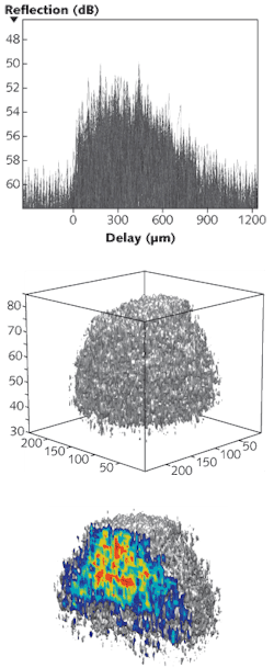
In the coherence-gating experimental setup, a low-coherence probe beam (from a 450 mW femtosecond Ti:sapphire laser) input from one arm of a modified Mach-Zehnder interferometer irradiates a tissue sample. Scattered light at different depths within the sample loses its coherence after a few scattering events, while a small portion of the beam is reflected coherently from inhomogeneities within the sample. These coherent “ballistic” photons have a different time of flight as they enter the recording medium, corresponding to a particular depth within the tissue sample. In PR coherence-gated holography, only those ballistic photons that retain coherence with a reference source give rise to a refractive-index modulation within the PR medium. Spatial heterodyning demodulates the image from the PR film onto a CCD sensor to create 2-D data slices that comprise the combined 3-D image (see figure).
Axial and lateral resolution of the HOCI PR method was reported as 12 and 10 µm, respectively, for tissue samples with wide fields of view and tissue depths up to 800 µm. The data acquisition rate of 8 × 10⁵ voxels/s means that 3-D scans at 800 µm tissue depth over an 800-µm-wide area would take approximately 16 s at a 25 mm step delay and a frame rate of 2 frames/s, says Michael Salvador, postdoctoral research assistant at the University of Cologne. “Our results represent an important step toward a reliable biomedical imaging tool using a PR polymer composite as the recording medium,” he says. “However, HOCI is not limited to biological tissue samples but might be applied in monitoring fabrication processes where the internal structure of transparent material is of paramount importance, such as the inner core of an optical fiber.” The researchers are exploring the use of superluminescent diodes as the light source.
REFERENCE
- M. Salvador et al., Optics Express 17(14) p. 11834 (July 6, 2009).
About the Author

Gail Overton
Senior Editor (2004-2020)
Gail has more than 30 years of engineering, marketing, product management, and editorial experience in the photonics and optical communications industry. Before joining the staff at Laser Focus World in 2004, she held many product management and product marketing roles in the fiber-optics industry, most notably at Hughes (El Segundo, CA), GTE Labs (Waltham, MA), Corning (Corning, NY), Photon Kinetics (Beaverton, OR), and Newport Corporation (Irvine, CA). During her marketing career, Gail published articles in WDM Solutions and Sensors magazine and traveled internationally to conduct product and sales training. Gail received her BS degree in physics, with an emphasis in optics, from San Diego State University in San Diego, CA in May 1986.