In 2008, researchers at the Max Planck Institute for Biophysical Chemistry (Göttingen, Germany) and the German Cancer Research Center (Heidelberg, Germany) developed an isotropic stimulated-emission-depletion (isoSTED) microscopy system capable of performing three-dimensional cell imaging with a resolution of 40 to 45 nm. Now, one year later, the same research group has developed a STED microscope that uses a single optical path for the excitation and STED beams, reducing mechanical drift associated with the conventional setups and significantly simplifying the microscope’s optical design.1
In STED microscopy, the (diffraction-limited) focal spot of the excitation beam is superimposed with the ring-shaped focus of the STED laser beam that, by virtue of stimulated emission, blocks fluorescence from occurring anywhere except from molecules located in the very small dark center of the STED focus. The effective focal-point size is thus reduced, resulting in an improved imaging resolution.
Simplifying alignment
Although the introduction of a single supercontinuum laser source (rather than a complicated titanium:sapphire master oscillator, regenerative amplifier, and optical parametric amplifier) has enabled the wider use of STED microscopy, the optical setup typically involves alignment of the excitation and STED beams in three directions as well as in time, requiring the operator to have experience in optical alignment.
To combat this complexity, the Max Planck research team devised a modified STED microscope that entirely eliminates one beam path and is aligned by design. The excitation and STED beams are coupled into a single polarization-preserving single-mode optical fiber. The beams exit the fiber and are focused to the same position in the sample, eliminating mechanical drift. The challenge for this single-beam, common-path STED microscope is the design of a phase filter that acts on the STED beam and leaves the excitation beam unaffected.
After determining that diffractive optical elements only operated in low-numerical-aperture setups and increased resolution by only 20%, and that spatial light modulators were complex and expensive, the researchers selected a phase filter based on the spectral dispersion properties of different optical materials. The phase filter consists of two optical media with refractive indices matched at the excitation wavelength of 625 to 635 nm, but notably different at the 750 nm STED wavelength. By bonding together four wedges of the alternating materials to create a modified vortex phase filter, a toroidal or donut-shaped point-spread function for the STED beam is obtained with a central round excitation beam.
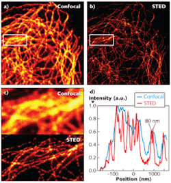
The single-beam STED setup was used to image a microtubular network of fluorescent-stained mammalian cells and the images were then compared to those obtained from a standard confocal microscope setup (see figure). The improved STED setup was able to resolve individual microtubules not evident with the confocal setup. “STED microscopy is often regarded as a rather complicated high-resolution microscopy method,” says Max Planck researcher Lars Kastrup. “Our latest results, however, emphasize that appropriate designs enable STED microscopes that are no more complex than common confocal instruments. Moreover, the single-path design introduces drift insensitivity as a strategic advantage.”
REFERENCE
- D. Wildanger et al., Optics Express 17(18) p. 16100 (Aug. 31, 2009).

Gail Overton | Senior Editor (2004-2020)
Gail has more than 30 years of engineering, marketing, product management, and editorial experience in the photonics and optical communications industry. Before joining the staff at Laser Focus World in 2004, she held many product management and product marketing roles in the fiber-optics industry, most notably at Hughes (El Segundo, CA), GTE Labs (Waltham, MA), Corning (Corning, NY), Photon Kinetics (Beaverton, OR), and Newport Corporation (Irvine, CA). During her marketing career, Gail published articles in WDM Solutions and Sensors magazine and traveled internationally to conduct product and sales training. Gail received her BS degree in physics, with an emphasis in optics, from San Diego State University in San Diego, CA in May 1986.
