CARS MICROSCOPY: NIVI quantitatively detects cancer cells

Diagnosing cancer in tissue biopsies has traditionally been done by experienced pathologists viewing a stained tissue sample under a microscope. They try to find cells that appear unusual in some way from the subcellular to the tissue level, often consulting their colleagues for confirmation. Besides being slow, taking a day or more, the technique is subjective.
Scientists at the University of Illinois at Urbana-Champaign have solved both these problems by developing an interferometric microscopy technique that is both fast and objective, explicitly outlining tumor boundaries in a biopsy in less than five minutes at a confidence level of more than 99%.1 Called nonlinear interferometric vibrational imaging (NIVI), the technique combines elements of optical coherence tomography (OCT) interferometric principles with coherent anti-Stokes Raman scattering (CARS). While maintaining the high throughput of CARS, NIVI uses interferometry to capture only coherent information, eliminating the nonresonant CARS background that limits quantitative analysis.
Both amplitude and phase measured
The laser system produces 100 fs broadband pump, Stokes, and reference pulses at 810, 1060, and 655 nm wavelengths, respectively. The pulses are focused to a spot in the tissue about 2 μm wide and 10 μm in the axial direction; the tissue sample is raster-scanned at a speed of 500 μm/s, with each scan line containing 100 pixels.
Both the amplitude and the phase of the CARS spectrum are acquired by mixing the CARS beam with a reference pulse time-delayed by 2 ps, with a grating and a camera capturing the spectral interferogram. An inverse Fourier transform is performed on the data, with the imaginary part providing the vibrational spectrum. The very sensitive nature of the NIVI measurements compensated for scattering losses in thick tissue samples (in the same way that the inteferometric measurements in OCT do).
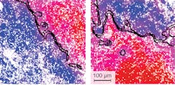
While normal cells have high concentrations of lipids, cancerous cells produce more protein, so by identifying cells with abnormally high protein concentrations, the researchers can accurately differentiate between tumors and healthy tissue. An edge-detection algorithm automatically finds the tumor margins, making sure that the margin is connected throughout the entire image (see figure).
Linearly proportional signal
Stephen Boppart, the head researcher, notes that NIVI was invented in his laboratory with the first theoretical publication in 2004.2 "The majority of my background work has been in OCT, and novel exogenous contrast agents for OCT," he says. "NIVI evolved as a new way for optical molecular imaging of endogenous species, by using OCT interferometry principles to detect the coherent signals from CARS processes."
In addition to the elimination of the nonresonant background, another important advantage of NIVI is that it provides a quantitative signal that is linearly proportional to the molecular concentration, he adds. "With these advantages, we can now clearly differentiate contributions of molecular species (collagen-proteins, and adipose-lipids, in this case), which can be used to characterize cells and regions within tissues. There have been several recent studies that link changes in the collagen-lipid ratio to breast cancer, and we use this for our margin detection, which turned out to be very sensitive and accurate."
The researchers plan next to further increase the spatial resolution so that they can probe subcellular molecular species. "We are already investigating skin specimens ex vivo, and plan to investigate many other tissue/tumor types," says Boppart. "Our goal here would be to identify early molecular cancer changes in cells/tissues before morphological/structural changes are evident on histology. And, related, we have been developing compact and portable fiber-laser sources that will enable NIVI to be performed with a portable system. We have already performed intraoperative OCT to identify positive tumor margins based on structural changes.3 Now, we intend to move NIVI into more clinical studies, performing intraoperative NIVI for this same application."
REFERENCES
1. P.D. Chowdary et al., Cancer Research, 70, 23 (Dec. 1, 2010).
2. D.L. Marks and S.A. Boppart, Phys. Rev. Lett., 92, 123905 (2004).
3. F.T. Nguyen et al., Cancer Research, 69, 8790-8796 (2009).

John Wallace | Senior Technical Editor (1998-2022)
John Wallace was with Laser Focus World for nearly 25 years, retiring in late June 2022. He obtained a bachelor's degree in mechanical engineering and physics at Rutgers University and a master's in optical engineering at the University of Rochester. Before becoming an editor, John worked as an engineer at RCA, Exxon, Eastman Kodak, and GCA Corporation.