ENDOSCOPY: Coherent Raman scanning-fiber endoscope images at 7 fps

Both stimulated Raman scattering (SRS) and coherent anti-Stokes Raman scattering (CARS) microscopy use light from a pump and probe laser beam scanned over tissue or a test sample to accomplish chemically selective, label-free, high-spatial-resolution imaging at up to video rates. These coherent Raman laser-scanning microscopes were not able to be miniaturized easily for endoscopy. But researchers at Harvard University (Cambridge, MA) and the University of Washington (Seattle, WA) have succeeded in developing a coherent Raman scanning-fiber endoscope, enabling SRS and CARS microscopy for in situ biological tissue analysis.1
In the standard coherent Raman scattering test setup, a 1064 nm neodymium:yttrium vanadate (Nd:YV04) laser provides 80 MHz repetition rate, 7 ps Stokes (probe-beam) pulses. A portion of this output is frequency doubled and sent to an optical parametric oscillator (OPO) to produce the pump beam at 800 nm. The Stokes beam is modulated and combined with the pump beam via a dichroic mirror. The beams are then linearly polarized and properly oriented for delivery along the proper axis of a 1-m-long section of polarization-maintaining (PM) optical fiber.
Spiral scanning
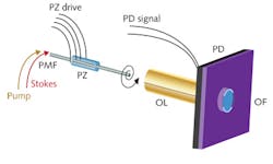
The fiber tip is driven at its mechanical resonance frequency in an expanding spiral by a piezoelectric actuator, enabling a scan rate of 7 frames per second (fps). The scan pattern is created by inserting the tip of the PM fiber through a 0.5-mm-diameter, piezoelectrically active ceramic tube radially patterned with four electrodes (see figure). With one pair of opposing electrodes driven with a sine wave and the other driven with a cosine wave (both with linearly expanding envelope functions), a spiral scan pattern is achieved. The driver electronics track the fiber tip for image reconstruction as the data is processed.
The researchers avoided the typical axial chromatic aberrations of a single-element gradient-index (GRIN) lens by using a chromatically corrected hybrid lens consisting of two GRIN lenses sandwiching a diffractive lens to focus the excitation light. The 1.4-mm-diameter lens uses the diffractive optic to achieve near-zero axial chromatic aberration at the chosen wavelengths. Because the sources emit picosecond pulses, ordinary silica fibers can be used for light delivery over 1 m without significant self-phase-modulation broadening of the optical spectrum beyond the intrinsic Raman linewidth (approximately 1 nm at 800 nm).
Experimental imaging of 2-µm-diameter polystyrene beads in an 80-µm-diameter field of view yielded axial full-width at half-maximum (FWHM) resolution of 6.5 µm and lateral FWHM resolution of 1.3 µm. Imaging of mouse tissue and hair confirmed the SRS scanning-fiber endoscope system was able to distinguish the chemical contrast between lipids and proteins at depths up to 30–50 µm into tissue. The contrast is comparable to previous CARS and SRS measurements. “There has been a lot of interest in developing miniaturized coherent Raman systems for medical applications in vivo, because the unique combination of label-free chemical contrast and microscopic resolution shows great promise for distinguishing healthy tissue from cancerous tumors, for example,” says Brian Saar, lead author of the paper and a graduate student at Harvard University when this work was done.
REFERENCE
1. B.G. Saar et al., Opt. Lett., 36, 13, 2396–2398 (2011).

Gail Overton | Senior Editor (2004-2020)
Gail has more than 30 years of engineering, marketing, product management, and editorial experience in the photonics and optical communications industry. Before joining the staff at Laser Focus World in 2004, she held many product management and product marketing roles in the fiber-optics industry, most notably at Hughes (El Segundo, CA), GTE Labs (Waltham, MA), Corning (Corning, NY), Photon Kinetics (Beaverton, OR), and Newport Corporation (Irvine, CA). During her marketing career, Gail published articles in WDM Solutions and Sensors magazine and traveled internationally to conduct product and sales training. Gail received her BS degree in physics, with an emphasis in optics, from San Diego State University in San Diego, CA in May 1986.