OPTICAL COHERENCE TOMOGRAPHY: Dual-window technique enhances true-color spectroscopic OCT
Spectroscopic optical coherence tomography (SOCT) and variations such as Raman spectroscopic OCT (RS-OCT) often use absorption contrast agents such as gold nanoparticles to enhance diagnostics; however, these particles require modification to be compatible with infrared (IR) illumination. The light sources can also limit analysis of many biological components that absorb in the visible region.
An alternative technique from Duke University (Durham, NC) researchers called molecular imaging true-color spectroscopic (METRiCS) OCT uses a broadband laser source (centered in the visible spectrum) and a dual-window (DW) data-processing method that improves imaging when using various IR and visible absorption contrast agents, simultaneously improving spectral and depth resolution compared to IR-only OCT systems.1
The experimental setup is based on a parallel Fourier-domain OCT (pfdOCT) system—essentially, a Michelson interferometer with an additional 4f imaging system that focuses a supercontinuum source (center wavelength 575 nm, bandwidth 240 nm) through a cylindrical lens and onto a beamsplitter to create an illumination line on the sample and the reference arm. Scattered light from the sample and reflected light from the reference arm enter a spectrograph that samples up to 400 interferograms (limited by CCD and beam size) in parallel with 1.2 μm axial and 6.9 μm transverse resolution.
Dual-window data and colorimetric contrast
The DW technique uses bilinear data processing to produce high-resolution spectral and spatial information. The DW algorithm computes two short-time Fourier transforms (STFTs), one with a wide spectral window and one with a narrow window, then multiplies the two resultant time-frequency distributions to form a single distribution that avoids the typical tradeoffs between spatial and spectral resolution that accompany single-STFT methods.
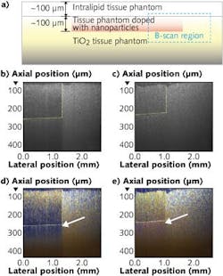
To demonstrate SOCT, gold nanosphere (GNS) and gold nanorod (GNR) contrast agents were first spectrally characterized through METRiCS OCT, revealing extinction peaks at 550 and 620 nm, respectively, in a deionized water/glycerol mixture. The GNR and GNS contrast agents were injected into layers of a tissue phantom—a physical model that represents nanoparticle-injected biological tissues (see figure).
The spectrum of each spatial point is extracted from the interferograms using the DW technique and then divided into RGB channels via the Commission Internationale d’Eclairage (CIE) color chart to be superimposed with the OCT image, producing a true-color or SOCT image. Analysis of various contrast agents and tissue samples can produce colorimetric and spectral contrast in three dimensions and discriminate nanoparticle concentrations to the sub-nanomolar level.
Using METRiCS OCT and DW data processing, the researchers have demonstrated true-color, quantitative, in vivo OCT imaging of rat dorsal skin in a windowed chamber and were also able to quantify vascular hemoglobin oxygen saturation levels.
You Leo Li, a research assistant in the Duke University BIOS laboratory, says, “We are planning on developing this approach for cancer imaging where the use of these contrast agents can provide cellular specificity and permit identification of cancer type.”
REFERENCE
1. Y.L. Li et al., Biomed. Opt. Expr., 3, 8, 1914–1923 (2012).

Gail Overton | Senior Editor (2004-2020)
Gail has more than 30 years of engineering, marketing, product management, and editorial experience in the photonics and optical communications industry. Before joining the staff at Laser Focus World in 2004, she held many product management and product marketing roles in the fiber-optics industry, most notably at Hughes (El Segundo, CA), GTE Labs (Waltham, MA), Corning (Corning, NY), Photon Kinetics (Beaverton, OR), and Newport Corporation (Irvine, CA). During her marketing career, Gail published articles in WDM Solutions and Sensors magazine and traveled internationally to conduct product and sales training. Gail received her BS degree in physics, with an emphasis in optics, from San Diego State University in San Diego, CA in May 1986.
