Raman Spectroscopy: Multi-wavelength excitation in Raman spectroscopy
GREGORY STAPLES, HUAWEN (OWEN) WU, and JACK QIAN
Thanks to photonics advances driven by the telecommunications boom of the 1990s, Raman spectroscopy has become much more accessible to users in all fields. In combination with its ability to perform nondestructive and in situ measurements of chemistry and structures with molecule-level sensitivity, the technological improvements have led to a surge of applications in such areas as pharmaceutics, biomedicine, and forensics, among others.1
A key parameter of a Raman system is excitation wavelength, which affects both depth of penetration into the sample (which is proportional to the laser wavelength) and Raman scattering intensity (inversely proportional to laser wavelength). While for many applications, a 532 nm or even 488 nm laser is the appropriate choice, organic samples can be highly fluorescent, with an emission about 1000x stronger than the Raman signal—which greatly reduces sensitivity. Fluorescence can be reduced or eliminated by selecting longer excitation wavelengths, such as 785 nm, though dark tissues such as kidney and liver exhibit extraordinarily high fluorescence and can benefit from even longer wavelengths. For instance, 1064 nm for excitation is even more effective for fluorescence elimination, but many commercial Raman systems use CCD detectors, which are insensitive beyond 1050 nm.
Ideally, a Raman spectrometer would have the ability to excite at different wavelengths to maximize the efficiency of different samples. Since this capability requires multiple lasers and multiple detectors, however, it has not been offered in compact systems. But thanks to new lasers, optics, and InGaAs detector technology (originally developed for the telecommunications industry), multi-wavelength excitation Raman analyzers have become available in handheld and benchtop form factors. Equipped with volume phase gratings (VPGs),2 such systems offer modular integration, compactness, high performance, low maintenance, and long-term reliability for meeting real-world challenges.3 Compared with the option of two separate systems, owning a multi-wavelength system is less expensive because of shared components, and offers additional benefits including a single user interface, shared Raman libraries and accessories, overlapping measurement volumes, and elimination of recalibration when switching between spectrometers.
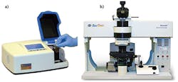
With such integration, accuracy is maximized under all conditions. In fact, multi-wavelength Raman is available in research-grade instrumentation, including a high-performance, three-wavelength (532, 785, and 1064 nm) Raman microscope; a transportable benchtop system (available in 532, 785 and 1064 nm in single- or dual-band models); and a scientific-grade dual-band Raman spectrometer (see Fig. 1).Application examples
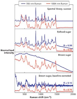
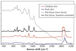
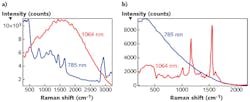
Because of the resolution and improved Raman scattering efficiency, library matching using 785 nm excitation is preferred for low-fluorescence materials. When fluorophores are present, however, significantly better Raman spectra can be obtained with longer-wavelength excitation. This fact is exemplified in measurements and signal-to-noise ratio (SNR) analysis of white and brown sugars (see Fig. 2). Since white sugar has lower fluorescence, excitation using 785 nm provides higher resolution Raman spectra at faster integration times. Dark sugars, however, contain such colored compounds as fluorescent furfurals, and require the use of a 1064 nm laser to avoid the noisy spectrum seen at 785 nm.As mentioned, pigmented and porphyrin-rich tissues such as kidneys are similarly too fluorescent to be measured at 785 nm. Using 1064 nm, a clear Raman spectrum, relatively devoid of fluorescence background, is generated using the same integration time. Additionally, because of the extended quantum efficiency of the InGaAs detector, high wavenumber features (C-H, O-H, and N-H stretching modes) are also simultaneously captured with the same laser. This is in contrast to high-wavenumber Raman on a 785 nm spectrometer, in which these bands present at the edge of silicon (Si) detectability, thereby necessitating a second, shorter wavelength laser source (usually 671 or 720 nm) and suitable filters and optics to accommodate both laser lines. Moreover, the high background signal of the glass at 785 nm pervades the measurement volume of the tissue and cannot be subtracted, leaving no resolvable Raman bands. At 1064 nm, this background signal is much reduced and can be simply subtracted to reveal clear Raman bands. The high spectral quality of a 1064 nm Raman system is expected to push biomedical research to a new frontier (see Fig. 4a).4
Plant biomass is another perfect example to demonstrate the advantages of 1064 nm Raman. Colorful plant samples are loaded with various photosynthetic pigments such as chlorophylls and carotenoids. Traditionally, plant researchers could not benefit from the advantages of Raman spectroscopy because samples fluoresce significantly and saturate the detector, and no visible laser is suitable. With 1064 nm Raman, a fluorescence-free, high-quality spectrum can be obtained almost in real time. This will significantly boost the analytical horsepower in the fields of plant biology, food science, and biofuels, among others (see Fig. 4b).
In another example, single-cell Raman spectroscopy enabled by confocal Raman microscopes offers a direct, non-destructive, quantitative in vivo diagnostic means for high-throughput analysis without requiring any exogenous modification of samples.5 Importantly, 1064 nm Raman brings significant benefit on biological cells or tissue samples, as these samples are often dyed (fluorescently labeled) for other assays before Raman measurement. Because a 1064 nm laser does not excite those fluorescence labels, their Raman spectra can be acquired (see image at top of page).
These examples demonstrate the power of Raman spectroscopy, which is an increasingly popular technique for life sciences because of its non-contact, non-destructive approach and ability to be applied in situ. Biological tissues have great variations in their fluorescence properties, which can limit the applicability of single laser excitation systems. This challenge is resolved with multi-wavelength systems, such as the portable Agility dual-band benchtop Raman spectrometer; the high-performance, scientific-grade dual-band RamSpec Raman spectrometer; and the high-performance, three-wavelength Nomadic Raman microscope. With the flexibility in design of VPGs, we can configure a system to match the varying requirements in real-world applications.
REFERENCES
1. J. Ferraro, Introductory Raman Spectroscopy, 2nd ed., Elsevier (2002).
2. S. Zhang and W. Yang, "Compact double-pass wavelength multiplexer-demultiplexer having an increased number of channels," U.S. Patent 6,108,471 (Aug. 2000).
3. W. Yang et al., "Multi-wavelength excitation Raman spectrometers and microscopes for measurements of real-world samples," Proc. SPIE, 8546: Optics and Photonics for Counterterrorism, Crime Fighting, and Defence VIII (2012).
4. C. A. Patil, I. J. Pence, C. A. Lieber, and A. Mahadevan-Jansen, Opt. Lett., 39, 2, 303–306 (2014).
5. H. Wu et al., Proc. Nat. Acad. Sci., 108, 9, 3809–3814 (2011).
Gregory Staples is sales manager for spectroscopy products, Huawen (Owen) Wu is an applications scientist, and Jack Qian is manager of application development, all at BaySpec, San Jose, CA; e-mail: [email protected]; http://www.bayspec.com.


