Photoacoustics/Multispectral Imaging: Optoacoustic/hyperspectral approach promises effective arthritis treatment
While chronic joint inflammation such as rheumatoid arthritis and osteoarthritis has no cure, when caught early it can be held in check with medication.
Early detection, though, has remained elusive: conventional x-ray imaging enables detection only at an advanced stage. Doppler ultrasound is a likely candidate, but detection at an early stage still remains challenging. Magnetic resonance imaging (MRI) enables detection earlier than x-ray, but is significantly more expensive than both of these techniques.
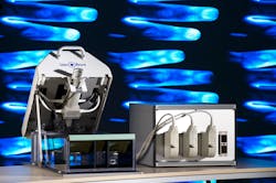
Now, a European consortium composed of several research institutions and companies, and led by Fraunhofer Institute for Biomedical Engineering (IBMT; Saarland, Germany), is developing an alternative technique combining ultrasound technology with light-based methods to enable very early diagnosis of arthritis in the hands (see figure). "Many forms of arthritis affect the fingers first," says Dr. Marc Fournelle, manager of the project (called IACOBUS) at Fraunhofer IBMT.
A 3D finger scanner searches joints for sites of inflammation and other pathological changes. It uses optoacoustic imaging, in which the fingers are subjected to extremely short laser light pulses of variable wavelength. As the tissue absorbs the light, the tissue expands, slightly causing faint pressure pulses that the scanner registers using an acoustic transducer—just as with ultrasound imaging. From the pattern of the pressure pulses, the device can pinpoint exactly where inflammation is forming.
To refine the diagnosis, the procedure also uses hyperspectral imaging, which scans the finger using strong white light and analyzes which wavelengths are absorbed by the inflamed tissue.
The system is able to generate ultrasound images using the scanner's acoustic transducer, which Fournelle explains is important because it gives physicians familiar output. The system then combines the ultrasound imagery with hyperspectral and optoacoustic data, enabling doctors to clearly identify inflammation sites.

Barbara Gefvert | Editor-in-Chief, BioOptics World (2008-2020)
Barbara G. Gefvert has been a science and technology editor and writer since 1987, and served as editor in chief on multiple publications, including Sensors magazine for nearly a decade.
