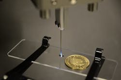
Low-cost two-photon-polymerization method could enable 3D printing of microstructures within the body
Two-photon polymerization using femtosecond lasers can be used to 3D-print detailed microstructures for research and medical purposes; however, femtosecond lasers are large and expensive. Now, scientists at École Polytechnique Fédérale de Lausanne in Switzerland have used an ultrathin multimode optical fiber to allow the use of an inexpensive continuous-wave (CW) laser emitting at a 488 nm wavelength as the light source for two-photon-polymerization 3D microprinting.1
The approach could one day be used with an endoscope to fabricate small biocompatible structures directly into tissue inside the body; this capability could enable new ways to repair tissue damage.
Phase-controlled multimode fiber
A 5-cm-long multimode fiber with a diameter of 70 μm and a numerical aperture (NA) of 0.64 is phase-controlled to produce a small focal spot near the fiber end; this plus the nonlinearity of the photopolymer allow the 3D printing. The approach can create microstructures with a 1.0 μm lateral (side-to-side) and 21.5 μm axial (depth) printing resolution. Although these microstructures were created on a microscope slide, the approach could be useful for studying how cells interact with various microstructures in animal models, which would help pave the way for endoscopic printing in people.
"With further development, our technique could enable endoscopic microfabrication tools that would be valuable during surgery," says research team leader Paul Delrot. "These tools could be used to print micro- or nanoscale 3D structures that facilitate the adhesion and growth of cells to create engineered tissue that restores damaged tissues."
By printing delicate details onto large parts, the new ultracompact microfabrication tool could also be a useful add-on to today's commercially available 3D printers that are used for everything from rapid prototyping to making personalized medical devices. "By using one printer head with a low resolution for the bulk parts and our device as a secondary printer head for the fine details, multi-resolution additive manufacturing could be achieved," says Delrot. "Compared to two-photon-photopolymerization state-of-the-art systems, our device has a coarser printing resolution; however, it is potentially sufficient to study cellular interactions and does not require bulky optical systems nor expensive pulsed lasers. Since our approach doesn't require complex optical components, it could be adapted to use with current endoscopic systems."
Moving toward clinical use
The researchers are working to develop biocompatible photopolymers and a compact photopolymer delivery system, which are necessary before the technique could be used in people. A faster scanning speed is also needed, but in cases where the instrument size is not critical, this limitation could be overcome by using a commercial endoscope instead of the ultrathin fiber. Finally, a technique to finalize and postprocess the printed structure inside the body is required to create microstructures with biomedical functions.
REFERENCE:
1. P. Delrot et al.,Optics Express, Volume 26, Issue 2, 1766-1778 (2018); doi: 10.1364/OE.26.001766.

John Wallace | Senior Technical Editor (1998-2022)
John Wallace was with Laser Focus World for nearly 25 years, retiring in late June 2022. He obtained a bachelor's degree in mechanical engineering and physics at Rutgers University and a master's in optical engineering at the University of Rochester. Before becoming an editor, John worked as an engineer at RCA, Exxon, Eastman Kodak, and GCA Corporation.
