Ongoing research using an existing imaging technique could provide a deeper understanding than ever before of neurological diseases such as Alzheimer’s and their impact on the brain. It could also pave the way for more effective treatment.
With a National Institutes of Health BRAIN Initiative grant award, a team at Boston University (BU) Neurophotonics Center—alongside researchers from the University of California San Diego, Massachusetts General Hospital (MGH), and the University of Illinois at Chicago—is leveraging neurophotonics to develop a technique for gathering information about neuronal circuits and their activity using functional MRI (fMRI), a noninvasive imaging method to measure and map brain activity. With this technique, researchers expect to get a better understanding of how the brain works and, specifically, how diseases disrupt normal brain functions.
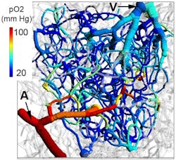
“In animals, it’s possible to do all sorts of invasive measurements that can be super specific,” says Dr. Anna Devor, a biomedical engineer (BME) and associate professor at BU leading this BRAIN Initiative award-supported study. “One example is two-photon phosphorescence lifetime microscopy (2PLM) in combination with oxygen-sensitive nanoprobes to map cerebral oxygen.” (see Fig. 1)
Development of this technology was led by Dr. Sava Sakadzic of the A. A. Martinos Center for Biomedical Imaging at MGH, a member of the BRAIN team. “2PLM has revolutionized our ability to observe oxygenation of brain tissue and vasculature,” Devor says. “With it we can calculate the rate of oxygen consumption by active neurons, which is a parameter needed to model fMRI signals. 2PLM requires that an oxygen-sensitive nanoprobe be delivered to the brain. In addition, it is necessary to open the skull for detection of phosphorescence.”
These are invasive procedures that obviously cannot be done on humans, she adds. So the team uses mice, where there is “a rich neurophotonics toolkit at our disposal,” including 2PLM, which was developed by Devor’s team members.
The advantage of fluorescence and phosphorescence imaging methods is their “exquisite specificity” for the involved brain cells and molecules, according to Devor. “You know exactly what it is you’re looking at.” Neurons generate electrical impulses, release neurotransmitters, consume oxygen, and regulate blood flow. With neurophotonic tools, each of these parameters can be addressed.
In contrast to these optical measurements, fMRI signals are more complex, reflecting blood flow dynamics and the balance between oxygen delivery and consumption. This is where 2PLM comes to the rescue. By studying neuronal activity together with its effects on blood flow and oxygenation, Devor’s team will test the limits of deriving specific information about neuronal circuits activity from macroscopic fMRI measurements.
Although fMRI measures neuronal activity indirectly, Devor notes this technology is “one of those amazing tools out there that’s used quite a lot in human research” due to its noninvasive nature. Its effectiveness in measuring and mapping brain activity was discovered about 20 years ago—scientists observed that when the amount of deoxygenated hemoglobin decreased due to the influx of fresh blood called upon by active neurons, the magnetic resonance signal increased.
“There’s a lot of interest to push this technique into the clinical setting for diagnosing disease, but that has been super hard because of an incomplete understanding of the relationship between activity of specific neuronal cell types, release of specific signaling molecules, and blood flow,” Devor says. “If we want one day to evaluate some drugs or treatments based on fMRI measurements, these measurements need to provide some information at the same level of cellular and molecular specificity.”
With the BRAIN Initiative support, Devor and her colleagues are studying the relationship between fMRI signals that fluctuate in time across the cerebral cortex and neuromodulatory inputs, projecting to the cortex from deep brain reconfiguring of local neuronal circuits.
Tracking brain activity
Neuromodulatory systems control behavioral states, such as the level of attention, arousal, and cognitive acuity. As they have a strong influence on behavior and cognitive ability, dysfunction of these systems in brain disease contributes to cognitive decline.
“Our natural brain activity is composed of an activity of local circuits, as if you have individual electrical components on a chip,” Devor says. “While each of the components is there to perform a certain function, we can interface with the chip and send some signals from outside, for example to bias voltages, or set the gain. We are also able to do this in our brain with those neuromodulatory systems.”
Among them is the cholinergic system, in which cholinergic neurons residing in a deep subcortical region called the basal forebrain project all over the cerebral cortex releasing a neurotransmitter acetylcholine, a signaling molecule that helps coordinate activity across brain regions and dials overall cognitive ability up and down. The cholinergic system “becomes dysfunctional early on at the onset of Alzheimer’s disease and other dementias leading to shortage of acetylcholine,” she says. “It would be very valuable to use noninvasive imaging to diagnose things like this. You could see that something is happening with your cholinergic activity in the brain, and that knowledge could guide your physician to come up with a treatment regimen that is right for you.”
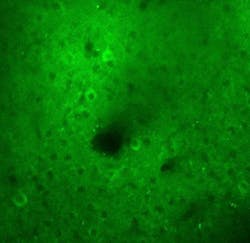
The team aims to ultimately use fMRI measurements to garner more precision in those neurotransmitter-specific systems. Parallel studies will be conducted in humans and animals. Starting with state-of-the-art in vivo multiphoton optical microscopy, the researchers will observe mice as they perform various tasks that require concentration, which will activate the release of acetylcholine (see Fig. 2), to see the relationships between neuronal circuit activity and blood flow. Some of these optical measurements will be done simultaneously with fMRI using high-field animal MRI scanners at the Martinos Center.In animals, researchers can use two-photon microscopy to look directly at the release of neurotransmitters such as acetylcholine, dilation of blood vessels, and consumption of oxygen. “From there, we can predict what our noninvasive fMRI signals are going to look like,” Devor says. “Then we can combine the optical measurements together with fMRI to confirm that our model works.”
The team will also perform parallel fMRI measurements in humans to ensure the model translates across scales and from mouse to human. To obtain human data, the researchers will combine fMRI with electroencephalography (EEG) and track other measures of alertness in humans, including the eye pupil diameter and skin conductivity.
“This would be very important in terms of basic science, and may have a clinical impact,” Devor says, noting the cholinergic system’s degeneration in Alzheimer’s disease. “This is an overall exploration of the human brain. We need to be able to interpret noninvasive human data in the language of the last 200 years of neuroscience knowledge that has been derived largely from animal studies. A bridge between the microscopic view of brain activity and that obtained with noninvasive technologies does not quite exist. We’re trying to bridge that gap.”

Justine Murphy | Multimedia Director, Digital Infrastructure
Justine Murphy is the multimedia director for Endeavor Business Media's Digital Infrastructure Group. She is a multiple award-winning writer and editor with more 20 years of experience in newspaper publishing as well as public relations, marketing, and communications. For nearly 10 years, she has covered all facets of the optics and photonics industry as an editor, writer, web news anchor, and podcast host for an internationally reaching magazine publishing company. Her work has earned accolades from the New England Press Association as well as the SIIA/Jesse H. Neal Awards. She received a B.A. from the Massachusetts College of Liberal Arts.

