Material made of fluorescent nanosensors may boost neural discoveries
The neuroscience community is always searching for ways to make measurements from neurons with the highest spatial and temporal resolution, to the extent possible with available technologies.
Dr. Abraham Beyene, a group leader at Howard Hughes Medical Institute’s Janelia Research Campus (Ashburn, VA), is creating highly sensitive biosensor solutions to address needs within the neuroscience research community.
In conjunction with Northern Ireland-based Raptor Photonics, Beyene’s team developed DopaFilm, a 2D composite material made of a layer of fluorescent nanosensors passivated by a second layer of polylysine (a natural homopolymer with a wide spectrum of antimicrobial activity with low toxicity to humans).
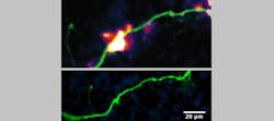
DopaFilm helps measure neurotransmitters released from neurons via fluorescent nanosensors that glow brighter when exposed to dopamine. According to research published in eLife, DopaFilm facilitates visualization of the release and diffusion of the neurochemical dopamine with synaptic resolution and quantal sensitivity, and simultaneously from hundreds of release sites.1
How it all works
Of course, monitoring the release of neurotransmitter chemicals is difficult to perform at spatial and temporal scales relevant to neuron function. Yet, improving the spatial and temporal resolutions of measurements for neurotransmitter release is crucial to fully understand chemical neurotransmission.
The researchers can monitor the spatiotemporal dynamics of dopamine release in dendritic (branched) processes with the DopaFilm technology. “What makes DopaFilm special is its ability to modulate (i.e., increase) its fluorescence brightness whenever it detects the neurotransmitter dopamine,” Beyene says.
Researchers use this property by growing dopamine-releasing neurons on top of the film, enabling detection of the release activity with a previously unachieved spatial resolution.
Whenever dopamine neurons cultured and growing on the DopaFilm fire electrical impulses and release dopamine, the area of the DopaFilm in contact with the part of the neuron releasing dopamine lights up, allowing the researchers to capture it at video frame rates with a microscope.
Raptor’s microscope is equipped with two fiber-coupled near-infrared (NIR) lasers—one operating at 671 nm and another at 785 nm—to excite far-red, NIR, and shortwave-infrared (SWIR) fluorophores, as well as a camera featuring optimized sensitivity in the SWIR range. It facilitates broad-spectrum imaging in the visible to SWIR ranges (400–1400 nm).
At the core of DopaFilm lies a fluorescent nanomaterial: single-wall carbon nanotubes (SWCNTs). SWCNTs are not only fluorescent, but also incredibly photostable and sensitive to their local chemical environment.
“This means you can make sensors out of them,” he says. “The dopamine sensor is based on SWCNTs. DopaFilm cleverly deploys these nanomaterials as a 2D film that affords a type of measurement that was not possible with existing tools and techniques.”
As the DopaFilm technology reveals precisely where the neurotransmitter originates and how it spreads between cells in real time, the researchers found that dopamine emerges a subset of dendritic processes as hot spots touting a mean spatial spread of roughly 3.2 µm at specific sites in cells. The researchers say these hotspots are observed with a mean spatial frequency of one hotspot per ~7.5 µm of dendritic length.
This technique is also helping the researchers better study dopamine’s release from subcellular compartments that previously were not well characterized.
Detectors featuring indium gallium arsenide (InGaAs) sensors—that operate in the SWIR and NIR ranges, commonly used in bio and life science applications—are needed for photon detection as well as additional optimization of optical components to facilitate transmission of photons in the SWIR and NIR ranges.
The new technology possesses non-photobleaching fluorescence properties in the NIR and SWIR regions of the electromagnetic spectrum (0.85–1.35 μm), making it spectrally compatible with existing optical technologies.
A notable challenge in sensing neurochemicals such as dopamine, which are released by neurons, is that these molecules are released into an extracellular place where they quickly diffuse or get cleared out by cellular processes.
“Sensing these molecules is a challenge because the sensor needs to be out in the extracellular space to capture these fleeting signals,” Beyene says. “Current approaches, which rely on protein-based sensors, anchor the sensors to the cell membrane and can provide only a partial readout and offer an incomplete picture. With DopaFilm, because it is 2D, it is a bit omnipresent and can recapitulate the spatial information fully, starting at the center of the release site, and following the signal to the outer edges.”
What’s next?
The researchers’ next steps will include tracking various biological questions, such as the characterization of the biophysical properties and protein machinery at dopamine (and other neurotransmitter) release sites. From a tool perspective, Beyene’s team is particularly interested in expanding its DopaFilm approach to sensing other types of neurotransmitters.
“The brain is an incredibly complex organ and there is still a lot to learn about it,” he says. “Tools and methods like DopaFilm will accelerate discoveries. Synthetic nanomaterials can make significant contributions to biomolecular sensing in the years to come. Most of the current sensing technologies rely on proteins and enzymes, and this class of nanoparticle-based sensor stands to make important contributions to biomedical research to augment the capabilities of existing tools.”
REFERENCE
1. C. Bulumulla et al., eLife, 11, e78773 (Jul. 4, 2022); https://doi.org/10.7554/eLife.78773.
About the Author
Justine Murphy
Multimedia Director, Digital Infrastructure
Justine Murphy is the multimedia director for Endeavor Business Media's Digital Infrastructure Group. She is a multiple award-winning writer and editor with more 20 years of experience in newspaper publishing as well as public relations, marketing, and communications. For nearly 10 years, she has covered all facets of the optics and photonics industry as an editor, writer, web news anchor, and podcast host for an internationally reaching magazine publishing company. Her work has earned accolades from the New England Press Association as well as the SIIA/Jesse H. Neal Awards. She received a B.A. from the Massachusetts College of Liberal Arts.

