Spectrally resolved fiber photometry system enables exploring the brain’s networks
A team at the University of North Carolina School of Medicine (Chapel Hill, NC) is working to crack the code of the brain’s default mode network (DMN)—a vast, very active, large group of regions linked to functions such as daydreaming and memory recollection, and problems with it are linked to diseases such as Alzheimer’s and attention deficit hyperactivity disorder (ADHD).
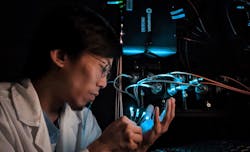
The researchers developed an experimental platform—currently being tested on rodent models—that involves neuroimaging and fiber optics (see video). It uses functional MRI (fMRI), a noninvasive imaging technique that measures blood flow changes in brain activity, as well as fiber photometry, which uses optical fiber to stimulate neurons with light and measure fluorescent signals relating to calcium dynamics (see Fig. 1).
During the past two decades, fMRI has emerged as the most prominent tool for noninvasive identification of large-scale functional networks such as the DMN and the salience network (SN). But noninvasive fMRI techniques have inherently low temporal resolution and are unable to resolve neuronal activity. It’s also been difficult to understand the neuronal mechanisms involved that govern the causal control of large-scale brain networks.
Spectrally resolved fiber photometry system
Their platform’s spectrally resolved fiber photometry system can obtain high-resolution emission spectra from multiple fluorescent sensors.
“This allows the use of an unmixing algorithm to extract each spectral component from the measured mixed spectrum,” Shih says, “and eliminates signal cross-contaminations caused by spectral bleed-through of multiple fluorescent sensors, which cannot be done with commercially available fiber photometry systems.”
Eliminating cross-contamination enables concurrent measurement via multiple fluorescent sensors with distinguishable emission spectra. Hemoglobin (Hb) in blood vessels absorbs light and affects optical measurement accuracy. It can have a significant impact on readout, so it’s important to quantify fluctuations in Hb concentration to ensure accurate photometry-based fluorescent sensor activity measurements. The team’s new platform is designed to do this.
“Our platform takes advantage of the fact that oxy- and deoxy-Hb absorb the fluorescent signal differently across wavelengths and use the dynamic distortions of the full emission spectrum shape from a stable fluorescent source to quantify the oxy- and deoxy-Hb concentration changes,” Shih says. “This information can then be used to correct Hb absorption artifacts in fiber photometry data.”
Quantifying fluorescent signals by fitting the whole spectra should also provide a better signal-to-noise ratio compared to quantifying the signals by the total photon counts within single bandwidth windows; for example, using a photomultiplier tube. “We are currently working on comparing these two kinds of systems in this regard,” Shih says.
This platform can also be adapted for the study of transfer functions between stimulation events, neuronal activity, neurotransmitter release, and hemodynamic responses.
Upgrading the system
Now, the team is upgrading the new system to record activity in three brain regions in two rodents simultaneously with multiple fluorescent sensors. This will allow them to investigate the DMN and SN roles in social interaction.
“Our multimodal approach will allow us to simultaneously test a number of critical hypotheses about macroscopic brain network function and organization,” Shih says. “Importantly, the framework and general principles we develop will set the stage for future investigations of large-scale, functionally and behaviorally significant brain networks in the healthy brain, and the neuronal mechanisms that lead to network dysfunction in brain disorders.”
He adds that this research has the potential to transform the fMRI landscape, and “the knowledge gained will have widespread implications for the design, analysis, and interpretation of human brain fMRI data.”
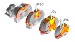
The team’s findings also reveal a neuronal basis of causal inhibitory control signals from the anterior insula, which regulates subjective feelings into cognitive and motivational processes, to the retrosplenial cortex (RSC; a cortical area in the brain that makes connections with numerous other brain regions), which facilitate access to attentional resources as observed in hemodynamic fMRI studies.
The team is expanding the study, as well, with the development of a silent zero echo time (ZTE) fMRI technique. It uses “auditory oddball paradigms” to manipulate the SN and DMN in awake, stress-free mice, Shih explains. The researchers are working to record excitatory and inhibitory neurons in the RSC, the anterior cingulate gyrus (Cg; a component of the limbic system that’s involved in processing emotions and behavior regulation), the prelimbic cortex (PrL; a protein hormone that controls reproduction, immunomodulation, and energy metabolism), and the anterior insula simultaneously during a silent fMRI sequence. Here, the researchers will also manipulate selected neuronal populations at specific brain regions to investigate the neuronal mechanisms of DMN and SN modulation in head-fixed awake mice.
“These methods, combined with computational modeling, have revealed critical in vivo evidence of the causal influence of direct anterior insula activation on SN-DMN dynamics,” Shih says, noting the team will expand upon their findings “with ground-truth neuronal information” using the new fiber photometry platform.
About the Author
Justine Murphy
Multimedia Director, Digital Infrastructure
Justine Murphy is the multimedia director for Endeavor Business Media's Digital Infrastructure Group. She is a multiple award-winning writer and editor with more 20 years of experience in newspaper publishing as well as public relations, marketing, and communications. For nearly 10 years, she has covered all facets of the optics and photonics industry as an editor, writer, web news anchor, and podcast host for an internationally reaching magazine publishing company. Her work has earned accolades from the New England Press Association as well as the SIIA/Jesse H. Neal Awards. She received a B.A. from the Massachusetts College of Liberal Arts.


