Advancements in laser tech impacting flow cytometry
The fundamental mechanism of fluorescence in flow cytometry relies on the interaction between fluorophores and incident light, which involves the absorption of photons and subsequent emission of light at longer wavelengths. Lasers are integral to this process, because of their photophysical and photochemical properties’ uniqueness as coherent and monochromatic light sources.
When a fluorophore absorbs a photon from the laser, it gets promoted from its ground electronic state to an excited state. This transition occurs through a process known as absorption, and the energy difference between these states corresponds to the energy of the absorbed photon. Following this, the fluorophore undergoes internal conversion and vibrational relaxation, leading to a loss of energy in the form of heat. Eventually, the fluorophore returns to its ground state through fluorescence emission, releasing a photon. This emitted photon, characterized by a longer wavelength and lower energy than the absorbed one, is detected and analyzed by the flow cytometer, which provides valuable information about the sample under study.
Choosing a laser over other light sources
It is essential to consider several key factors when selecting a suitable light source for optimal performance. By understanding the implications of spectral width, intensity, and stability, researchers can make informed decisions that significantly impact the quality and reliability of their flow cytometry data.
The first step should be considering the chosen fluorophore excitation spectrum. The choice of laser wavelength must be carefully matched to the excitation spectrum of the fluorophore under study, ensuring efficient and selective excitation.
The laser light source’s spectral width is also crucial. Unlike a LED with a wide spectral width, the laser possesses a narrower spectral width that can minimize the risk of unwanted cross-excitation of spectrally adjacent fluorophores, which is crucial for multiplexed experiments that require simultaneous detection of multiple fluorophores. Coherence is directly related to and inversely proportional to spectral width. High spatial and temporal coherence increases excitation efficiency, provides more effective signal collection, and reduces background noise. However, excessive coherence can result in the formation of speckle patterns and interference effects, which might negatively impact the accuracy of flow cytometry data—a balancing act.
Power stability is essential, as well, to ensure reproducibility and consistency across experiments. Fluctuations in laser output power or wavelength can introduce variability (a.k.a. errors) into flow cytometry measurements, potentially confounding data interpretation and jeopardizing the accuracy of the results. Selecting a light source with excellent long-term power stability and low noise characteristics is vital, ensuring that the data generated remains reliable and comparable across multiple experiments and time points.
Both tunable and supercontinuum lasers are often proposed as light sources in flow cytometry. Tunable lasers provide the flexibility to adjust the excitation wavelength, optimizing the laser light source for the specific fluorophores. On the other hand, supercontinuum lasers generate a broad spectrum of wavelengths, enabling simultaneous excitation of multiple fluorophores with varying excitation profiles. Both tunable and supercontinuum lasers facilitate advanced multiplexing and offer greater adaptability for various applications—but they are infrequently viewed as optimal tools in flow cytometry.
For instance, tunable and supercontinuum lasers may introduce increased cytometer design and alignment complexity, requiring specialized expertise to maintain and operate. And these units can cost upward of four to five times the price of a discrete laser source, leading many to turn to multiple discrete lasers to accomplish their different fluorophores of inquisition. These upscale lasers also often have power limitations that eliminate many fluorophores from consideration.
The development of micro-LEDs and micro-lasers requires a delicate balance between miniaturization and performance, ensuring that the reduced size does not compromise the quality, power production, or reliability of flow cytometry measurements. As the field of flow cytometry continues to evolve, researchers must carefully consider the advantages and limitations of these emerging photonics technologies to harness their potential in further driving flow cytometry innovation fully. As it stands today, micro-LED technology in its current form yields output power that is too low, and true microscale lasers may be nearly a decade away from being stable enough for research purposes.
Laser performance
The performance of lasers in flow cytometry is governed by several interconnected factors that, when optimized, contribute to the efficient excitation of fluorophores and accurate data collection. First, beam quality ensures a consistent and well-defined excitation profile. An optimal beam quality, characterized by a low M2 value and high spatial coherence, allows for a tightly focused laser spot at the interrogation point, improving signal resolution and sensitivity.
Wavelength stability is also essential, because it directly influences the excitation and detection of specific fluorophores. A stable wavelength ensures reliable and reproducible results, minimizing deviations in acquired data that may occur due to fluctuations in the laser’s output wavelength. This characteristic is particularly crucial when using multiple lasers or fluorophores with narrow excitation bands.
Laser power must also be considered, because it directly affects the signal-to-noise ratio, allowing for the detection of dimmer signals and enhanced separation of distinct cell populations. Excessive laser power may result in photobleaching and phototoxicity, however, compromising sample integrity and cell viability. Balancing these factors is essential to achieve accurate and reliable flow cytometry results.
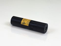
For example, Power Technology’s instrument-quality IQ and CK series of laser diode modules are designed explicitly to address the needs of clinical research and flow cytometry applications that require superior optical quality and ultra-stable temperatures, wavelengths, and output powers. These lasers feature a precision operating current source and a PID temperature control loop, allowing for beam-pointing stability less than 5 μRad/°C. These modules feature onboard microprocessors, as well, providing advanced user control and monitoring capabilities.
Applications in clinical research
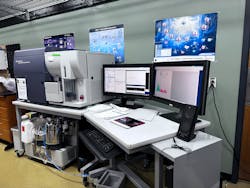
The multifaceted realm of flow cytometry has become a powerful analytical technique with a wide array of applications in clinical research. Advances in laser technology are driving the increasing sophistication of cytometer instrumentation.
For instance, protein and biomarker detection remains a cornerstone of flow cytometry applications, facilitating the identification and quantification of cellular components and signaling molecules. This approach enables researchers to unravel the complex interplay of cellular processes and uncover potential therapeutic targets.
Flow cytometry has become instrumental in the development and execution of cell-based assays, contributing to our understanding of cellular responses to stimuli and evaluating potential drug candidates. Researchers can obtain valuable insights into drug efficacy and safety by monitoring parameters such as cell viability, proliferation, and apoptosis.
Flow cytometry also excels at microbial analysis, providing rapid and accurate identification, enumeration, and characterization of diverse microbial populations.
The technique has revolutionized our ability to study and manipulate these complex ecosystems, from assessing bacterial viability to monitoring changes in microbial community structure. Flow cytometry continues to break new ground in myriad other applications—from stem cell research and immunophenotyping to the study of intracellular signaling pathways and beyond. As laser technology and flow cytometry instrumentation continue to evolve, there is little doubt this versatile technique will continue to play a pivotal role in shaping the future of scientific discovery.
Lasers vs. other light sources
When considering efficiency in fluorophore excitation, lasers have a clear edge over alternative light sources, such as arc lamps and LEDs. The high-intensity, monochromatic light emitted by lasers provides superior excitation of fluorophores, leading to increased sensitivity and specificity in flow cytometry measurements. This is particularly important when working with low-abundance targets or multi-color applications that require the simultaneous excitation of several fluorophores.
Lasers typically have a narrow spectral width, minimizing spectral overlap between fluorophores and reducing background noise. In contrast, arc lamps and LEDs often have broader emission spectra, which can result in increased crosstalk between fluorophores and decreased sensitivity. But recent advancements in LED technology have led to the development of narrow-bandwidth LEDs, which may soon offer improved spectral characteristics compared to traditional LEDs—still a far cry from what lasers can offer. Along with their unmatched stability, this is why lasers are the gold standard for quality data collection in flow cytometry.
Impact of laser technology on flow cytometry
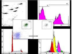
Lasers enable the development of more sensitive and precise methods for analyzing complex samples. Using lasers with stable, narrow spectral widths and high intensities provides the simultaneous detection of multiple fluorophores, ultimately increasing the multiplexing capabilities of flow cytometry assays. As a result, researchers can now obtain detailed information on numerous cellular parameters in a single experiment, significantly improving the efficiency and depth of analysis.
The impact of lasers on flow cytometry is particularly evident in cancer research and drug discovery, where advanced cytometers have enabled the exploration of new applications. For instance, laser-driven flow cytometry is instrumental in characterizing cancer stem cells, which play a crucial role in tumor growth and metastasis. And the high-throughput capabilities of flow cytometry, bolstered by laser technology, have facilitated large-scale screening of compound libraries for drug discovery efforts.
Along with these advancements, laser technology is helping develop novel flow cytometry techniques such as time-resolved fluorescence, which can provide valuable insights into the dynamic behavior of cellular processes. This further extends the applicability of flow cytometry in biomedical research, allowing for more comprehensive investigations into cell signaling, gene expression, and protein-protein interactions.


