Near-infrared light enables optical observation in microscopy
Imaging with fluorescent proteins, dyes, and quantum dots can help visualize biological events, and visible light often serves as the excitation source. But there are several challenges when using visible light. Its high energy can cause fluorescence photobleaching and phototoxicity to the biological specimen, and it has a limited penetration depth due to scattering and absorption in specimens and autofluorescence.
To mitigate challenges associated with using visible light as an excitation source, researchers are turning to near-infrared (NIR) imaging. It is currently used in a variety of biological research applications, and new NIR fluorescence probes are being developed to expand what’s possible with fluorescence imaging. Likewise, microscope manufacturers are also adding new tools to their microscope systems to enable NIR imaging.
Multiphoton excitation
Multiphoton excitation (MPE) takes advantage of NIR using high-power pulse lasers. It’s a well-established technique for optical sectioning used widely during in vivo imaging because it can penetrate farther into the sample to enable researchers to observe events that occur within deeper sample layers. NIR light has lower photon energy, which reduces issues associated with phototoxicity.
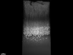
If two or more photons excite a fluorophore at the same time, it is excited with the same energy as a single photon and emits corresponding fluorescence. Even though the system uses NIR lasers, which have a longer wavelength than visible light, the light emitted is within the shorter wavelength visible spectrum. For example, green fluorescent protein (GFP) can be excited at 920 nm, but emits fluorescence around 500 nm. The multiphoton absorption of the fluorophore described above only happens in high-density photon areas, which correspond to the microscope’s objective focus spot. Therefore, the MPE image has optical sectioning and good contrast at various resolutions and allows researchers to observe events within living animals 1 mm+ beneath the sample surface (see Fig. 1).
MPE microscopes can also be used to make observations without fluorescence labeling. For example, second-harmonic generation (SHG) and third-harmonic generation (THG) imaging can be carried out via MPE microscope.
SHG is the optical process where two photons with the same frequency interact with nonlinear materials to produce twice the energy of the original photons. It enables researchers to observe collagen and other fibrous proteins without requiring labeling. This information can be combined with data captured using fluorescence for a more comprehensive picture of a sample. When using 920-nm excitation, SHG emits 460-nm light, which makes it easy to separate the emission light from GFP and SHG.
NIR fluorescence
Another area of innovation is NIR probes. Recently, a number of new fluorescence dyes that absorb NIR light and emit longer-wavelength light have come to market. They are gaining in popularity as microscope manufacturers add NIR light sources and detectors to their systems—which enables the use of NIR dyes. For example, 735-nm LED light sources and 685-nm, 730-nm, and 785-nm lasers excite NIR fluorescence dyes while new detectors, such as cameras and photomultipliers, are designed to handle a broader range of wavelengths from visible to NIR.
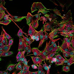
This setup provides a solution to the above-mentioned challenges, but also enables researchers to image one or two additional fluorophores without crosstalk—the unintended detection of fluorescence signals from one fluorophore in the channel designated from another fluorophore—which enables easier multiplex imaging (see Fig. 2).
Recently, many novel fluorescent dyes and fluorescence proteins were developed for increasingly longer wavelengths. As they continue to gain popularity, more NIR fluorescence dyes and proteins will undoubtedly come to market.
Upconversion
Another novel technology in probes is using upconversion, which is a process where longer-wavelength (that is, lower energy) photons are absorbed and re-emitted as shorter-wavelength (higher energy) photons. Upconversion is used in various fields, and solar cells are the most famous application. The sun emits visible light as well as IR and NIR light, but nonvisible light is not absorbed efficiently by solar cells. Upconversion helps convert NIR/IR light to visible light to improve solar cells’ efficiency.
In upconversion, lower-energy photons are converted into higher-energy photons through various mechanisms. One is called excited state absorption, and involves three steps:
1. A photon is absorbed by an ion, such as Er3+ or Nd3+), which excites it from the ground state to a higher energy state (first excited state).
2. The ion in the first excited state absorbs another photon and moves to an even higher excited state.
3. The ion in the higher excited state emits a photon with higher energy (shorter wavelength) than the absorbed photons.
This mechanism is interesting because it is similar to MPE. This mechanism was recently used in microscope imaging, and next-generation fluorescent biomarkers are being developed based on the technology. Unlike MPE, upconversion does not require that the probes be excited simultaneously, so it does not require a pulsed laser and can be accomplished using continuous-wave (CW) lasers.
Upconversion is also being studied for medical applications, such as diagnosing drug-induced liver injuries. Sensitive and real-time in vivo detection of hepatotoxicity is currently a bottleneck in the diagnosis of drug-induced liver injury.
In a paper published in 2020 by Professor Li Huijun's research group, an upconverting nanoprobe was designed by assembling upconversion nanoparticles and gold nanorods. It was then used to diagnose drug-induced liver injuries in vivo and in real time. This novel nanoprobe aggregates in the liver and is activated by a liver injury marker, miR122, producing fluorescence images at 800 nm when being excited by NIR light at 980 nm. In combination with luminescence resonance energy transfer and signal-amplification technology, its detection sensitivity is further increased to achieve highly sensitive detection of miR122—which provides a new approach for real-time clinical monitoring of drug-induced liver injury.1
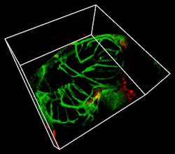
Furthermore, upconversion is also being explored as a cancer treatment because NIR light can penetrate deeper into tissue and activate photosensitizers, which generate oxygen species to attack cancer cells (see Fig. 3).2
NIR light is becoming increasingly popular and varied new applications are rapidly being developed. There are many ways to use the NIR wavelength for biology and life science applications, and its potential for future research and discovery is exciting.
REFERENCES
1. L. Meng et al., Nanoscale, 12, 28, 15325–15335 (Jul. 23, 2020).
2. R. Tian et al., Chem. Sci., 10, 10106–10112 (2019).
About the Author
Akira Saito
Akira Saito is manager of Advanced Imaging Solutions at Evident (Tokyo, Japan).


