Photon-counting detectors are poised to revolutionize computed tomography
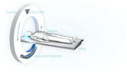
During a CT scan, three-dimensional (3D) images of the body are created using x-rays. For this purpose, an x-ray tube rotates around the patient (see Fig. 1), taking several images of a body region or organ from different angles. A 3D image is then generated by a computer, which a practitioner can use to assess an injury or look for signs of illness.
Since the 1970s, CT has been used in patient care and is one of the most essential tools used by radiologists. Although the technology has been used successfully for many years, research within the field of CT remains important. Numerous innovations have refined the procedure. The area of the detectors has been significantly increased, for example, to be able to scan larger parts of the body or entire organs, such as the heart, in one rotation. The time required for a scan was also significantly reduced to simultaneously reduce the impact of motion artifacts and increase the comfort for the individual patient. Recently, the goal of improvements has been to minimize radiation exposure for patients.
X-rays are ionizing and, in high doses, can even lead to long-term effects such as cancer. As such, reducing the dose administered to patients is a societal challenge driving many of the recent technological advances in CT. Furthermore, using as low a dose as reasonably achievable for a given diagnostic task is of utmost importance in pediatrics and cancer screening applications.
For about 20 years, researchers around the world have been working to create a new method to implement the next revolution in CT, which should mitigate more than just the problem of radiation exposure. And today, photon counting is on the verge of market maturity.
From indirect to direct detection
In conventional CT, indirect detectors are used. X-rays hit a scintillator layer and are converted into less energetic light, which can then be detected with photodiodes. The output signal of these photodiodes is proportional to the intensity of the incident x-rays.
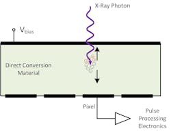
Photon counting, on the other hand, is based on a fundamentally different detection principle: direct-conversion semiconductors (such as cadmium telluride or cadmium zinc telluride) are used to convert individual x-ray photons directly into an electrical charge (see Fig. 2). Electrons drift toward the anode and induce a charge pulse proportional to the energy of the impinging photon. The signal generation is also fast enough so that the pulse processing electronics can distinguish individual photons and count them.
This makes the detector significantly more sensitive than in conventional CT. Due to the higher sensitivity and the fact that all photons contribute equally to the image, radiation exposure can be reduced by 40 to 80%, depending on clinical protocol. Photon counting also achieves better spatial resolution and higher contrast, which allows practitioners to obtain more and more reliable information from the images (see Fig. 3).
But photon counting has yet another advantage: it can also provide spectral information. Since each pulse also contains energy information, the front-end not only counts how many photons passed through the patient but also quantifies their energy and assigns each event to a limited number of energy bins (e.g., 5 bins). This is why CT with a photon-counting detector is also referred to as “spectral CT.” In the subsequent image pre-processing stage, the additional information enables separating the main mechanisms of x-ray interaction with matter, which allows quantification and identification of the tissue responsible for the attenuation within the body.
Just the beginning
The benefits outlined above are available to clinical practices with the adoption of photon counting. This technology, however, offers many other potential benefits likely to be introduced during the next decade.
One such example is “k-edge” imaging. Due to the nature of the technology, which allows counting and estimation of the energy of photons, it is possible to distinguish and quantify materials that present a discontinuity in their attenuation properties (a.k.a. “k-edge”). The introduction of contrast agents based on such materials will allow targeted diagnostics like the quantification of plaque, a severe risk factor in heart infarction.
Extensive research of these and other applications of CT with photon counting are currently underway. Although the introduction of such advanced imaging techniques will take significant time due to drug development, regulatory, and safety procedures, it does show that photon counting will sustain a high level of clinical advancements for the foreseeable future.
The spectral information is automatically obtained during photon counting and stored with the data. This means that even if it was not clear during an initial examination that spectral information was needed, it is immediately available if the need arises as the treatment progresses. This saves the patient a further examination and the associated radiation exposure.
Future outlook
Computerized tomography is one of the most important imaging techniques in modern medicine. Photon counting is poised to revolutionize this technology by enabling higher resolution and higher contrast while also reducing radiation exposure.
The first CT scanners with photon counting have been on the market since late 2021, and it is now up to semiconductor manufacturers to make the technology more readily available. With its photon-counting detectors, ams OSRAM plans to assist its customers on the journey to the next CT revolution.
About the Author
Roger Steadman
Roger Steadman is head of System Solution Engineering and systems architect in Computed Tomography for ams OSRAM.


