Two-photon microscope cracks neural networks code
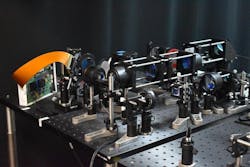
Researchers from the University of California, Davis developed a two-photon fluorescence microscope to capture high-speed images of neural activity at cellular resolution (see video).
The team’s design uses a short line of light that only illuminates specific parts of the brain where neurons are active. Unlike what’s possible with a single point of light, use of a short line allows larger areas to be sampled simultaneously, which significantly speeds up the imaging process. Another key advantage of the new microscope setup lies in not imaging the background and inactive areas—this reduces the total light energy needed, as well as potential damage to brain tissue.
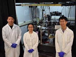
“This advancement is crucial because it allows scientists to monitor brain activity more faithfully in time and more safely, providing a clearer view of how neurons communicate in real time,” says Weijian Yang, an associate professor in the Department of Electrical and Computer Engineering.
Digital micromirror
The short point of light to illuminate specific areas of the brain involves adaptive sampling, a method in which accruing data and observations are used to adjust current research and experiments in real time.
For their work, the researchers used a digital micromirror—a device that contains thousands of tiny mirrors they can control individually—to shape and steer the light beams and precisely target the active neurons of interest. They can turn individual pixels in the device on and off, which allows them to adjust their approach toward the brain’s neuronal structure while it is imaged.
The team also used the digital micromirror to mimic high-resolution point scanning. Images can be reconstructed from the rapid scans, which facilitates higher-speed imaging and identifies neuronal regions of interest quicker.
Notable findings
An increase in imaging throughput to monitor brain activity via conventional two-photon microscopy has great implications in neuroscience. However, such increases in throughput typically mean increasing laser power. This can cause phototoxicity—a condition in which the skin or eyes become very sensitive to sunlight or other forms of light—in the brain.
To avoid it, Yang says the research team designed their microscope to be compatible with typical laser-scanning two-photon microscopy.
Through a series of experiments, they found their technology can also be easily integrated into a conventional microscope, too, using a galvo-galvo scanner, which uses two galvo scan mirrors to deflect a laser beam, or a resonant-galvo scanner, a type of galvanometric mirror scanner that allows fast image acquisition with single-point scanning microscopes.
Next steps
“Our ultimate goal is to provide a powerful and practical imaging tool for the neuroscience community to study the brain,” Yang says.
Accomplishing this will involve voltage imaging; specifically, integrating its imaging capabilities with the new microscope. With voltage imaging, electrical signals across neuron membranes can be measured. This produces a direct and extremely rapid readout of neural activity. Incorporating this technique will enable the observation of transient electrical changes in neurons with even greater temporal precision, and complement the calcium imaging that is currently used.
“Broadening the scope of our research will also include enhancing our microscope’s capabilities for volumetric imaging with the synchronization of binary mask changes on the digital micromirror device,” Yang says.
This will involve capturing 3D images of neural activity over time, which is crucial for understanding the complex spatial arrangements and interactions within neural networks. Implementing remote focusing techniques and faster scanning methods will be necessary to achieve efficient high-resolution volumetric imaging.
Another goal: applying these techniques in real-world neuroscience applications.
“Our current work provides a proof of demonstration of the technology,” Yang says. “But we would like to apply it in real neuroscience applications, as well. This could be to observe neural activity during learning, to study how and what the communication and interaction among different neurons evokes during this process, and to look more closely at brain activity during disease states.”
The future
The team prides itself on their two-photon fluorescence microscope design and development, because it represents a significant advancement in neural imaging technology. Yang says the technology, which enables high-speed imaging of neural activity with minimal photodamage, “opens new possibilities for studying dynamic neural processes with longer times.”
But there are even wider implications for its use in neuroscience and beyond, including an enhanced understanding of brain function overall. “Our technology allows researchers to explore aspects of brain function that were previously inaccessible,” Yang says. And it could boost scientists’ understanding of fundamental neurological processes.
It could help improve research into neurological diseases such as Alzheimer’s, Parkinson’s, autism, and epilepsy, in which it’s crucial to recognize changes in neural activity. This would lead to earlier detection of disease, better monitoring of disease progression, and ultimately more targeted therapies.
“The principles and technology developed for this microscope could also be adapted for use in other scientific fields that require precise imaging capabilities,” Yang says, citing developmental biology, as well as nonbiological fields where dynamic processes occur at microscopic scales.
FURTHER READING
Y. Li et al., Optica, 11, 1138–1145 (2024); https://doi.org/10.1364/optica.529930.
About the Author
Justine Murphy
Multimedia Director, Digital Infrastructure
Justine Murphy is the multimedia director for Endeavor Business Media's Digital Infrastructure Group. She is a multiple award-winning writer and editor with more 20 years of experience in newspaper publishing as well as public relations, marketing, and communications. For nearly 10 years, she has covered all facets of the optics and photonics industry as an editor, writer, web news anchor, and podcast host for an internationally reaching magazine publishing company. Her work has earned accolades from the New England Press Association as well as the SIIA/Jesse H. Neal Awards. She received a B.A. from the Massachusetts College of Liberal Arts.


