Photon-counting cameras for the life sciences
Since Robert Hooke and Jan van Leeuwenhoek made their first studies of microorganisms with early microscopes, optical imaging has been at the forefront of advancing knowledge of life and organic materials. A key challenge within the life sciences is imaging photosensitive samples that can easily experience photobleaching, where the strength of the signal dims due to overexposure, and photo damage, where the sample itself is harmed. These effects are prevalent in fluorescence microscopy, which typically use high-intensity laser sources for illumination to gain sufficient signal.
To minimize deleterious effects on photosensitive samples, life scientists illuminate them with low levels of optical power. But this leads to extremely faint fluorescence signals that are very difficult to detect without being swamped by the camera’s inherent noise, as well as being limited by the bounds of classical imaging such as shot noise.
This is where photon-counting cameras come in—they provide sufficiently low noise so that single-photon events can be registered on each individual pixel of the array. It makes them ideal for imaging dim samples with a tight “photon budget.”
Bridging the gap between novel camera technology and application to life sciences is not necessarily straightforward—there are different camera architectures available, with numerous user-controlled parameters to be considered and optimized.
The Centre of Light for Life (based in Adelaide, Australia) is well positioned to perform an analysis of cutting-edge camera technologies by combining expertise with sensitive biological samples and extremely “gentle” low-photodose microscopy techniques such as light sheet microscopy. The results of this research were conducted between the groups of Kishan Dholakia and Kylie Dunning and published as a tutorial in APL Photonics.1
Photon-counting cameras
Digital photography originated in the 1970s with the invention of the charge-coupled device (CCD) array. Advancements such as the development of complementary metal-oxide semiconductor (CMOS) arrays led to such refinement that the ability to snap a picture is now available on every smartphone. Scientists have benefited from the advancement of this technology, and these sensors can detect and register events right down at the single-photon level.
Detection of single photons is only possible due to the exceedingly low electronic noise of modern cameras. Quantum theory gives us only statistical predictions, and therefore it’s extremely difficult to distinguish between a photon and a bit of noise, so it must be kept to a minimum.
Combating electronic noise, much of which is thermal in nature, makes it necessary to cool camera sensors well below the freezing point of water. Further, instruments such as the electron-multiplying CCD (EMCCD) use what is effectively an internal photomultiplier to reduce electronic noise further—at the expense of introducing “excess noise” from the multiplication process.
Within the past few years the CMOS camera architectures have advanced to being worthy of scientific use. Scientific CMOS (sCMOS) cameras are steadily growing in popularity and sophistication, and the recently released “quantitative CMOS” (qCMOS) architecture of digital camera claims to offer similar photon-detecting capability to EMCCD cameras without the burden of excess noise.
Comparing CCD and sCMOS architectures, there are numerous minor tradeoffs regarding detection efficiency, pixel size, frame-capture rate, and unique sources of noise. While much of this is well understood and familiar to instrumentalists in the case of, say, astronomy, this is not true for the life sciences, which carry a whole host of different challenges (especially in regard to the range of sensible exposure times). Close analysis is necessary to get the most out of these cutting-edge technologies.
A boon for life sciences
Cameras are enormously powerful pieces of technology—images contain vast amounts of information and allow unprecedented insights into the natural world. Cameras are so common they can easily be taken for granted, and it’s tempting to adopt a “point and shoot” mindset when snapping pictures. But to be fully rigorous it’s necessary to think about every step of the imaging process—from the sample, through the optical system, onto the camera sensor, and into a computer hard drive. Every step in this process can introduce artefacts that distort physical reality, and it’s important to be mindful of this in the age of artificial intelligence (AI), where algorithms that only know what we give them are becoming increasingly dominant.
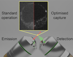
For instance, a major impetus behind our tutorial was to determine whether EMCCD or qCMOS cameras are better suited for quantitative imaging. To make this comparison fair, it was necessary to develop a procedure to determine signal-to-noise and contrast-to-noise ratios for fluorescent images in a way that was independent of pixel size or camera imaging settings. With this in hand, we discovered that the excess noise from the electron-multiplication process in EMCCDs inflated the effect of a fluorescence background, a persistent fact of fluorescence microscopy due to the incoherent nature of the emission.
Certain modern sCMOS/qCMOS cameras include a “confocal linescan mode” which helps to reduce this fluorescent background even further. For quantitative imaging, which may require an accurate biological measurement of relative intensity level, such as is the case for the study of certain metabolic factors, the qCMOS is the better choice. But the presence of row- and column-based fixed-pattern noise in the images can degrade certain datasets, especially those for training AI models. EMCCD cameras remain the most sensitive cameras available, and are still a good option when qualitative image analysis is of interest, such as imaging an extremely weakly fluorescent signal to confirm its presence.
What’s next?
The increasing advance of camera technology has a role to play in the “second quantum revolution.” For the life sciences, the ability to detect single photons allows another layer of physical understanding to be applied to images captured by microscopes. This could have powerful implications for advanced modalities such as super-resolution, single-molecule tracking, computational imaging, and quantum approaches such as ghost or sub-shot noise imaging. It means researchers must be extremely familiar with the cameras they use to get the most out of them and effectively apply them to the most challenging biological questions of our day, while illuminating the sample as gently as possible—imaging with a light touch.
REFERENCE
- Z. Peterkovic et al., APL Photonics, 10, 3, 031102 (Mar. 2025); https://doi.org/10.1063/5.0245239.
About the Author
Zane Peterkovic
Zane Peterkovic is a Ph.D. student at the Centre of Light for Life, Institute for Photonics and Advanced Sensing, University of Adelaide (Australia); www.adelaide.edu.au/centre-of-light-for-life and www.adelaide.edu.au/ipas.

