Microscope alteration on path to enhancing bioimaging
A new microscope prototype is taking standard ptychography to new depths, achieving the highest-ever numerical aperture and imaging throughput that is orders of magnitude greater than others previously demonstrated.
“This will enable novel optical instruments with inherent quantitative nature and metrological versatility,” says Guoan Zheng, the UTC associate professor in the University of Connecticut’s (UConn) Department of Biomedical Engineering.
The new technique, “coded ptychography,” was developed by Zheng’s team and builds upon ptychographic microscopy—a method of microscopic imaging in which a computer algorithm inverts an extensive data set (comprising numerous inference patterns obtained as an object’s position is shifted) into an image. Standard ptychographic microscopy is a coherent diffraction imaging technique for both fundamental and applied sciences, but is currently not as well suited for optical microscopy. According to the UConn team’s study, published in ACS Photonics, its applications in optical microscopy fall short due to the low imaging throughput and limited resolution.
In a conventional microscope setup, the objective lens is large and bulky. Also, looking at a sample with a regular objective lens often means a tradeoff between the resolution and imaging field of view.
“The motivation for us to develop this device is to try to get both,” Zheng says. “So now, we get high resolution and ultra-large field of view at the same time.”
For the coded ptychography prototype platform, the researchers essentially created their own imaging system by replacing the bulky objective lens by a very thin scattering layer. This allowed them to translate samples across disorder-engineered surfaces for lensless diffraction data acquisition. This surface consists of chemically etched, micron-level phase scatters and printed subwavelength intensity absorbers “for achieving the highest numerical aperture among all existing ptychographic systems.”
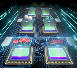
Figure 1 shows the scheme of the coded ptychography prototype, where the cover glass of the image sensor is replaced by the disorder-engineered surface. The engineered surface redirects large-angle diffracted light into smaller angles for detection. Therefore, the otherwise inaccessible object details can now be acquired using the pixel array.
According to the study, the resolution and numerical aperture the team has achieved is at least two times higher than current lensless ptychographic systems. “The unique fabrication process also differs from that of the conventional metasurface,” Zheng says, “allowing cost-effective manufacturing without involving optical or electron lithography.”
The engineered surface is fabricated on a microscope coverslip. When attached to the image sensor, the engineered facet faces the pixel array of the sensor. This arrangement avoids direct contact between external objects and the scattered layer. It also addresses the stability and alignment issues of a conventional scattering lens.
Zheng notes theirs is a computational objective lens, not a physical one, establishing a “resolution-enhanced parallel coded ptychography technique.” The researchers say their prototype is “designed to unlock an optical space with spatial extent and frequency content that is inaccessible using conventional lens-based optics.”
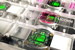
Lens-based systems often endure demanding requirements of mechanical stability. Images can be easily defocused due to misalignment or environmental vibrations. In the new ptychography device, the team overcomes this by encoding an empty region on the engineered surface for positional tracking. The device can operate without feedback from the mechanical stage, enabling open-loop optical acquisition. Figure 2 shows the prototype device developed by the UConn team.The researchers have been working on the coherent diffraction imaging model by considering both the spatial and angular responses of the pixel readouts. “Our low-cost prototype can directly resolve a 308 nm line width on the resolution target without aperture synthesizing. Gigapixel high-resolution microscopic images with a 240 mm2 effective field of view can be acquired in 15 seconds,” Zheng says.
They also demonstrated the ability to recover slow-varying 3D phase objects with many 2π wraps, including an optical prism, a convex lens, and bacterial colonies. “The low-frequency phase contents of these objects are challenging to obtain using other existing lensless techniques,” he adds.
Coded ptychographic microscopy in action
The technology is useful for several biomedical applications—among them, digital pathology. Coded ptychography can acquire high-resolution, whole-slide images of tissue sections for diagnosis.
“Typically, in the hospital, we take the biopsy sample, smear it on top of a glass slide, and then look at it under the microscope,” Zheng says. “We need to scan this sample at different positions to understand its overall structure. Our device, with its very large field of view, allows us to image the entire sample at once.”
Different from the conventional whole-slide scanner, Zheng says his team can perform post-acquisition digital refocusing of 3D thick samples. This capability also allows them to measure the profile of the lens and monitor the 3D shape of bacterial colonies on the Petri dish.
For regular light microscope, it’s challenging to set up 8 microscopes for parallel operation. “In our case, we don't have any objective lens, we only have 8 coded image sensors. In our study, we operate 8 sensors all at the same time. By using our platform, we can have 8 times higher throughput than whatever you can do using a single sensor,” Zheng says.
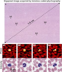
Blood screening is another potential application for coded ptychographic microscopy. For example, a gigapixel image acquired via this technique can be used for differential white blood cell counting, and in resource-limited settings, the new platform can be used to locate malaria parasites in blood smears. Figure 3a shows the recovered gigapixel image of a blood smear using the prototype platform. Figure 3b shows the zoomed-in views of the recovered phase and intensity images of white blood cells.For culture-based experiments in research or clinical laboratories, the new technology can quantify the cell cultures over the entire Petri dish. Mammalian cell lines at different wells can be monitored, and drug screening can be performed. Clinicians can also monitor bacterial growth in response to different concentrations or different types of antibiotics.
“We try to look at the bacteria’s image in response to different types of antibiotic drugs,” he says. “If you have the correct antibiotic shot, you can see immediately the bacteria’s cells quit dividing. But you’ll see growth of bacteria over time if that shot is not effective. So, we use this coded ptychography for drug screening to determine which antibiotic is the most effective.”
Currently, this can be done in a hospital by looking at the size of the bacterial colony in a Petri dish. But because Zheng’s team has been able to detect single-cell bacteria, they can quickly determine which cell is dividing and which is not.
Zheng notes the team is continuing to study such applications, alongside UConn John Dempsey Hospital, before the new technology can be used in clinical settings.
Outside of the bio industry, coded ptychography could also find applications in optical metrology, because the 3D shapes and surface profiles of various optical elements can be precisely measured.
What’s ahead?
The UConn team previously studied other super-resolution imaging techniques such as Fourier ptychography, where they illuminated a sample with different incident angles for resolution improvement.
“Our ongoing effort is to perform angle-varied illumination on this coded ptychography platform. By doing so we can further improve the resolution via aperture synthesizing and perform tomographic imaging of thick specimens,” Zheng says. “We will report these results soon.”
Ptychography is an enabling technique that has become an indispensable imaging tool in most synchrotron laboratories worldwide. Its application in high-throughput optical imaging, currently in its early stage, will continue to grow and expand. The UConn team’s research and findings are among the first steps in this direction.
About the Author
Justine Murphy
Multimedia Director, Digital Infrastructure
Justine Murphy is the multimedia director for Endeavor Business Media's Digital Infrastructure Group. She is a multiple award-winning writer and editor with more 20 years of experience in newspaper publishing as well as public relations, marketing, and communications. For nearly 10 years, she has covered all facets of the optics and photonics industry as an editor, writer, web news anchor, and podcast host for an internationally reaching magazine publishing company. Her work has earned accolades from the New England Press Association as well as the SIIA/Jesse H. Neal Awards. She received a B.A. from the Massachusetts College of Liberal Arts.


