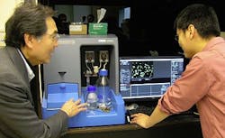
Nanoparticle tracking helps characterize 'nanoconstructs' for biomedical applications
Scientists at Duke University (Durham, NC)'s Fitzpatrick Institute for Photonics, led by Professor Tuan Vo-Dinh, applied nanoparticle tracking analysis (NTA) to characterize metal nanoparticle construct materials for use in biosensing, imaging, and cancer therapy.
To accomplish this, Dr. Hsiangkuo Yuan and other members of Vo-Dinh's group first designed and fabricated metal nanoparticle constructs such as gold nanostar platforms, which were then characterized with UV-VIS, transmission electron microscopy (TEM), Raman microscopy, fluorometers, and other techniques. But to design nanoconstructs for in-vivo applications, the particle size needs to be in the 10-100 nm range for lower clearance from the kidney and reticuloendothelial system (RES). It is also important that the construct is physiologically stable (non-aggregated) for biomedical applications such as optical imaging or nanodrug delivery, where the nanoparticle dose administered must be determined. To compare plasmonic properties (the enhanced electromagnetic properties of nanoparticles), they need to determine the effect of different sizes and to understand in detail the profile of the particle size distribution of similar concentrations. Recognizing this, the team turned to NTA (using an NTA system from NanoSight [Salisbury, England]).
Prior to NTA, the group mostly used TEM to look at particle shape and measure particle size. The surface coating or the aggregation state cannot be easily investigated using just TEM; NTA paired with TEM provides hydrodynamic size distribution and zeta potential. It also allows them to normalize their comparison by individual particle counting.
The team has published its work in the journal Nanotechnology, with another paper currently in press for Nanomedicine. For more information, please visit http://iopscience.iop.org/0957-4484/23/7/075102.
-----
Subscribe now to Laser Focus World magazine; it's free!
