NEAR-INFRARED FLUORESCENCE: Fluorescence promises earlier diagnosis, better treatment for osteoarthritis

A new study is reportedly the first to demonstrate that near-infrared (NIR) fluorescence can be used to monitor changes in osteoarthritis, a painful joint disease that is often detected only after agonizing symptoms occur. The approach might may make it easier to diagnose the disease—which could lead to better management and patient outcomes—and to help evaluate drugs for treatment.
The study, by researchers at Tufts University School of Medicine (TUSM; Boston, MA) and the Sackler School of Graduate Biomedical Sciences at Tufts, reports that a NIR fluorescent probe tracked activity leading to cartilage loss (the key characteristic of osteoarthritis) in the joints of male mice, brightening as the disease progressed.1
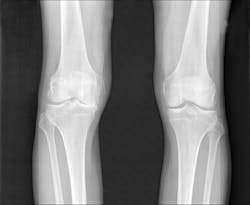
"The imaging tests most frequently used, x-rays, don't indicate the level of pain or allow us to directly see the amount of cartilage loss, which is a challenge for physicians and patients," says Averi A. Leahy, BA, an MD/PhD student in the medical scientist training program at TUSM and the Sackler School. "The fluorescent probe made it easy to see the activities that lead to cartilage breakdown in the initial and moderate stages," says Shadi A. Esfahani, MD, MPH, postdoctoral fellow in the division of nuclear medicine and molecular imaging at Massachusetts General Hospital, and in the department of radiology at Harvard Medical School.
The right knees of 54 mice were affected by injury-induced osteoarthritis and served as the experimental group (the mice received pain medication). The healthy, left knees served as the control group. Over two months, the researchers imaged each knee every two weeks. Strikingly, the signal became brighter in the injured right knee at every examined time point. The probe emitted a lower signal in the healthy left knee, and did not increase significantly over time.
Li Zeng, Ph.D., TUSM associate professor, reports that the next step is to monitor the fluorescent probe over a longer time period to determine whether the same results are produced during the late stages of osteoarthritis. She also hopes to use the probe to help develop treatments for animals.
1. A. A. Leahy et al., Arthritis Rheum., 67, 2, doi:10.1002/art.38957 (2015).
About the Author

Barbara Gefvert
Editor-in-Chief, BioOptics World (2008-2020)
Barbara G. Gefvert has been a science and technology editor and writer since 1987, and served as editor in chief on multiple publications, including Sensors magazine for nearly a decade.