SINGLE-MOLECULE MICROSCOPY/PORTABLE PATHOLOGY: Compact devices compete with high-end instruments
One compact device converts an ordinary smartphone into a fluorescence microscope capable of single-molecule detection and measurement, while another uses holography to do the work of large pathology lab microscopes. Both are new innovations from the lab of Aydogan Ozcan at the University of California, Los Angeles (UCLA).
Single molecule-capable smartphone
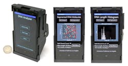
UCLA researchers reported the first demonstration of imaging and measuring the size of individual DNA molecules using a smartphone.1 The inexpensive, 3D-printed optical add-on uses a camera phone to visualize and measure the length of single-molecule DNA strands. An attachment to the device creates a high-contrast, dark-field imaging setup using an inexpensive external lens, thin-film interference filters, a miniature dovetail stage, and a laser diode that obliquely excites the fluorescently labeled DNA.
Labeled molecules are stretched on disposable chips that fit in the smartphone attachment. An app connects the smartphone to a server at UCLA; it transmits raw images to the server, which quickly measures the length of each DNA strand. The detection and measurement results can be seen on the mobile phone and on remote computers linked to the server.
Sizing accuracy is better than 1 kilobase-pair (kbp) for 10 kbp and longer DNA samples imaged over a ~2 mm2 field of view. The innovation holds promise for myriad applications for point-of-care medicine and global health.
Compact, lens-free pathology
The lens-free microscope works by using a laser or LED to illuminate tissue or blood on a slide that is inserted into the device.2 A cellphone-camera-style sensor array on a microchip captures and records the pattern of shadows created by the sample. The device processes these patterns as a series of holograms, forming 3D images of the specimen that provide a virtual depth-of-field view. An algorithm color-codes the reconstructed images, making the contrasts in the samples more apparent than they would be in the holograms, and highlighting abnormalities for easier detection.The invention could lead to inexpensive, portable technology for performing common examinations of biomedical specimens. It may prove especially useful in remote areas and in cases where large numbers of samples need to be examined quickly. Because it produces images several hundred times larger in area (field of view) than do conventional bright-field optical microscopes, it enables faster processing of specimens.
In a blind test, a board-certified pathologist analyzed sets of specimen images that had been created by the lens-free technology and by conventional microscopes. The pathologist's diagnoses using the lens-free microscopic images proved accurate 99 percent of the time.
Accompanied by advances in its graphical-user interface, the platform could scale up for use in clinical, biomedical, scientific, educational, and citizen-science applications, among others, says Ozcan.
1. Q. Wei et al., ACS Nano, doi:10.1021/nn505821y (2014).
2. A. Greenbaum et al., Sci. Transl. Med., doi:10.1126/scitranslmed.3009850 (2014).

Barbara Gefvert | Editor-in-Chief, BioOptics World (2008-2020)
Barbara G. Gefvert has been a science and technology editor and writer since 1987, and served as editor in chief on multiple publications, including Sensors magazine for nearly a decade.

