IN-VIVO IMAGING/MICROSCOPY: Portable microscope images individual blood cells noninvasively

Given the right equipment, shining light through the skin can reveal the same information that a traditional blood test yields-but in real time. The new optical microscopy device that enables the analysis is about the size of a shoebox. It provides high-resolution images of individual blood cells within veins-sans fluorescent dyes. The microscope promises not only to eliminate long wait times for blood test results, but it also might help spotlight warning signs (i.e., high white blood cell count) before a patient develops severe medical problems. And its size could enable doctors in rural areas to screen large populations for common blood disorders, notes Lior Golan, a graduate student in the biomedical engineering department at the Israel Institute of Technology (Technion; Haifa, Israel), where the device was developed.1
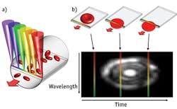
The device uses spectrally encoded confocal microscopy (SECM), which creates images by splitting a light beam into its constituent colors. A probe is pressed against the skin of a patient and the line of light is directed across a blood vessel near the surface of the skin. As blood cells flowing through the vessel cross the line, they scatter light, which is collected and analyzed. The color of this light carries spatial information, and computer programs interpret the signal over time to create 2D images of the blood cells.

The technique takes advantage of the one-way flow of cells to create a compact probe that can quickly image large numbers of cells while remaining stationary against the skin. The researchers used their invention to image blood flowing through a vessel in the lower lip of a volunteer. They then measured the average diameter of the red and white blood cells and also calculated the percent volume of the different cell types—which is key for many medical diagnoses.
A second-generation system with higher penetration depth might expand the range of possible imaging sites beyond the inside lip (which was selected because it is rich in blood vessels, has no pigment to block light, and doesn't lose blood flow in trauma patients). The team hopes to produce a thumb-size prototype within the next year.
1. D. Golan et al., Biomed. Opt. Exp., 3, 6, 1455–1464 (2012).
More BioOptics World Current Issue Articles
More BioOptics World Archives Issue Articles