IMAGE-GUIDED SURGERY/FLUORESCENCE: Tool illuminates low-grade tumors
In a pilot study, David Roberts, chief of neurosurgery at Dartmouth-Hitchcock Medical Center (Lebanon, NH), operated on 10 patients with gliomas using a new tool designed to highlight the tumors. Following the operations, a pathologist evaluated how accurately the probe had identified tumor tissue. The results are striking: Diagnoses based on fluorescence were accurate 64% of the time, but with the probe accuracy increased to 94%.1
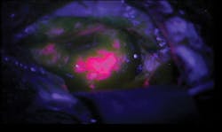
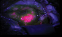
The probe, which boosts fluorescence highlighting for tumors that are less metabolically active, represents a collaborative effort between the Norris Cotton Cancer Center and Dartmouth's Thayer School of Engineering. When the researchers first saw the results of using their fluorescing agent and probe on low-grade brain tumors, it was "jaw-dropping. The tumor glowed like lava," according to Keith Paulsen, professor of engineering.
The patients participating in the study had been given oral doses of the chemical 5-aminolevulinic acid (ALA) prior to their surgery. When metabolized, ALA produces the protein protoporphyrin IX (PpIX), which accumulates faster in tumor cells than healthy cells—and makes cancerous tissue fluoresce when exposed to blue light. Throughout the surgeries, Roberts used a microscope to see the fluorescence, and the new handheld probe to evaluate sections where fluorescence was not definitive.
The probe combines violet-blue and white light to simultaneously analyze both the concentration of PpIX and four other tumor biomarkers: PpIX breakdown products, oxygen saturation, hemoglobin concentration, and irregularity of cell shape and size. The probe reads how light travels when it hits the tissue, and sends this data to a computer. The computer runs it through an algorithm, and produces a straightforward answer as to whether the tissue is cancerous.
The Thayer/Norris Cotton/Dartmouth-Hitchcock team, which has been working on the brain probe for about six years in collaboration with scientists from the University of Toronto, built on research conducted in Germany about 15 years ago.
The brain probe may have applications for other cancers as well; a protocol is already in place for a similar study on lung cancer tumors.
1. P.A. Valdes et al., J. Biomed. Opt., 16, 11, 116007 (2012).

