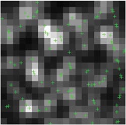
Using compressed sensing, researchers have boosted the spatial and temporal resolution of super-resolution microscopy techniques. The work makes super-resolution techniques more capable for live-cell imaging, as it enables resolving cell features an order of magnitude smaller than what could be seen before, and allows observation of single-cell structures in motion.1
Super-resolution microscopy methods such as stochastic optical reconstruction microscopy (STORM) and photoactivated localization microscopy (PALM) rely on the recording of light emission from a single molecule in the specimen. Using probe molecules that can be switched on and off, these approaches determine the position of each molecule of interest to define a structure. The single-molecule images are spread sparsely into many, often thousands of, camera frames. The process is limited in its temporal resolution and does not enable following of dynamic processes in live cells.
Compressive sensing is a novel sampling theory: It predicts that sparse signals and images can be reconstructed from what was previously believed to be incomplete information.2 "We can now use our discovery using super-resolution microscopy with seconds or even sub-second temporal resolution for a large field of view to follow many more dynamic cellular processes," says Lei Zhu of the Georgia Institute of Technology (Atlanta, GA), who, with Bo Huang and other colleagues at the University of California, San Francisco, conducted the research.
Using the same optical system and detector as in conventional light microscopy, super-resolution microscopy naturally requires longer acquisition time to obtain more spatial information, leading to a trade-off between its spatial and temporal resolution. In methods based on STORM/PALM, each camera image samples a very sparse subset of probe molecules in the sample. Increasing the density of activated fluorophores lets each camera frame sample more molecules—but high density causes fluorescent spots to overlap, invalidating the single-molecule localization method.
A number of reported methods can efficiently retrieve single-molecule positions, even when fluorophore signals overlap. These methods are based on fitting clusters of overlapped spots with a variable number of point-spread functions (PSFs), with either maximum likelihood estimation or Bayesian statistics. The Bayesian method has also been applied to the whole image set.
The new approach is based on global optimization using compressed sensing, which does not involve estimating or assuming the number of molecules in the image. They show that compressed sensing can work with much higher molecule densities compared to other technologies and demonstrate live-cell imaging of fluorescent protein-labeled microtubules with 3 s temporal resolution.
The authors' STORM experiment, with immunostained microtubules in Drosophila melanogaster S2 cells, demonstrated that nearby microtubules not discernible by the single-molecule fitting method can be resolved by compressed sensing using as few as 100 camera frames. The researchers have also performed live STORM on S2 cells stably expressing tubulin fused to mEos2.
At the commonly used camera frame rate of 56.4 Hz, the scientists constructed a super-resolution movie with a time resolution of 3 s (169 frames) and a Nyquist resolution of 60 nm, much faster than previously reported. These results prove that compressed sensing can enable STORM to monitor live cellular processes with second-scale time resolution, or even sub-second-scale resolution if a faster camera can be used.
1. L. Zhu, W. Zhang, E. Elnatan, and B. Huang, Nat. Meth., doi:10.1038/nmeth.1978 (2012).
2. M. Fornasier and H. Rauhut, Compressive Sensing, retrieved from http://rauhut.ins.uni-bonn.de/CSFornasierRauhut.pdf.
More BioOptics World Current Issue Articles
More BioOptics World Archives Issue Articles
