CELL BIOLOGY/IMAGING/ANALYSIS: Cell Bio event highlights and foretells change made possible by biophotonics

A session on quantitative live-cell microscopy by researchers from Harvard Medical School—presented during the American Society for Cell Biology (ASCB) annual meeting (December 3–7, 2011)—demonstrated how computer vision and biology are changing each other. Change, driven by both research need and technology innovation, was evident at the event, which participants call simply “Cell Bio.” So were at least four themes: Advanced image capture, multimodal microscopy, automation, and image analysis.
New high-performance camera systems exemplify the first theme: Hamamatsu promoted its new ORCA-Flash4.0 second-generation scientific CMOS (sCMOS) system, which combines a large field of view and low noise along with sensitivity, quantum efficiency, resolution, dynamic range, and speed. Along similar lines, Photonis introduced its xSCELL digital scientific camera designed for high-speed, low-light imaging with minimal noise.
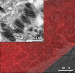
FEI Company’s new MAPS (Modular Automated Processing System) product illustrates the latter three themes, as it imports and correlates imagery from any type or brand of light microscope with that derived from an FEI scanning electron microscope (SEM) or DualBeam (focused ion beam/SEM) system. MAPS aims to facilitate quick navigation to a region of interest identified in the light image and leveraging of electron microscopy’s resolving power to get to the ultrastructural detail.
EMD Millipore used the ASCB event to launch its Muse Cell Analyzer, a small-footprint device that competes with imaging technologies by using laser-based fluorescence to evaluate up to three cellular parameters and provide accurate, quantitative results automatically.
Technology applied
The conference featured an array of presentations highlighting optical technologies, notably an evening talk by Jennifer Lippincott-Schwartz of the National Institutes of Health that packed an auditorium. Dr. Lippincott-Schwartz showcased the impact of recent developments in probes, techniques, microscopes, and quantification—including new photo-activatable fluorescent proteins (PA-FPs) and single, molecule-based super-resolution (SR) imaging—describing how such tools are helping researchers better understand the cell in order to answer long-standing questions.
Finally, in tutorial/exhibitor showcase presentations, vendors provided useful information on the application of various technologies for specific areas of cell biology. For instance, in separate sessions, Asylum Research and Bruker Corp. discussed the advantages of atomic force microscopy (AFM) for cell studies, Agilent explored the utility of laser combiners for advanced microscopy, and Carl Zeiss Microscopy highlighted the possibilities available by combining light and 3-D electron microscopy. In addition, both Accuri/BD Biosciences and TTP LabTech Ltd. described innovations in flow cytometry.

Barbara Gefvert | Editor-in-Chief, BioOptics World (2008-2020)
Barbara G. Gefvert has been a science and technology editor and writer since 1987, and served as editor in chief on multiple publications, including Sensors magazine for nearly a decade.