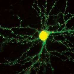
FLUORESCENT PROBES/NEUROSCIENCE: Fluorescing live synapses shed light on learning, memory formation
A new type of imaging probe is enabling real-time visualization of excitatory and inhibitory synapses that change as new memories are formed.1 The probes attach fluorescent markers to synaptic proteins, and light up synapses in living neurons without affecting neuronal functioning. In photomicrographs, the synapses appear as bright spots along dendrites (the branches that transmit electrochemical signals). As the brain processes new information, the bright spots change, visually indicating how synaptic structures in the brain have been altered by the new data.
"When you make a memory or learn something, there's a physical change in the brain," explains Don Arnold, associate professor of molecular and computational biology at the University of Southern California (USC; Los Angeles, CA), who co-led the work. "It turns out that the thing that gets changed is the distribution of synaptic connections."
To make these probes, the team used a technique known as "mRNA display," developed by Nobel laureate Jack Szostak along with chemistry and chemical engineering professor Richard Roberts. "Using mRNA display, we can search through more than a trillion different potential proteins simultaneously to find the one protein that binds the target the best," said Roberts, who led the team along with Arnold.
The probes, which the researchers call "FingRs," are attached to green fluorescent protein (GFP), which glows brightly when exposed to blue light. While they act like antibodies, they bind more tightly and are optimized to work inside the cell—something ordinary antibodies cannot do.
Because FingRs are proteins, the genes encoding them can be put into brain cells in living animals, causing the cells themselves to manufacture the probes (in fact, the probes can be put in the brains of living mice and imaged through cranial windows using two-photon microscopy). A regulation system cuts off the amount of FingR-GFP that is generated after 100% of the target protein is labeled, effectively eliminating background fluorescence and generating a sharper, clearer picture.
The researchers say that their work may offer crucial insight for scientists responding to President Barack Obama's Brain Research Through Advancing Innovative Neurotechnologies (BRAIN) initiative, which aims to help mankind better understand how we think, learn, and remember.
1. G.G. Gross et al., Neuron, 78, 6, 971–985 (2013).
