LIVE CELL IMAGING/MICROSCOPY: Low-intensity laser enables label-free approach to computed 3D live cell imaging
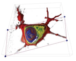
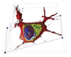
A pair of Swiss researchers has designed a device, based on holographic microscopy and computational image processing, that can create 3D images of living cells and track their reaction to stimuli without labeling.1 Their setup generates three-dimensional images with less than 100 nm resolution in just a few minutes; they are currently working to develop instantaneous operation. Because the approach requires no contrast dyes or fluorescent probes, foreign substances cannot impact experiment results. In addition, their use of a low-intensity laser minimizes the impact of light or heat on cells and enables observation over extended periods of time.
As the laser scans the specimen from various angles, a digital camera captures the images extracted by holography. The scans are then assembled by computer into a 3D image, and "deconvoluted" to eliminate noise. The assembled image can be virtually "sliced" to display cell components such as nucleus, genes and organelles.
The researchers think that being able to capture a living cell from every angle in this way opens the door to a whole new field of exploration. "We can observe in real time the reaction of a cell that is subjected to any kind of stimulus," explained Yann Cotte, who, with fellow researcher partner Fatih Toy, was lead author on a paper explaining the work.1 This approach would allow the study of pharmaceuticals at the scale of the individual cell, for example. Christian Depeursinge of the Microvision and Microdiagnostics Group at École Polytechnique Féderale de Lausanne (EPFL)'s School of Engineering supervised their work.
Toy and Cotte are launching a company, and in collaboration with Lyncée SA (also of Lausanne), they hope to develop portable systems able to generate in vivo scans. Their paper in Nature Photonics is accompanied by a time-lapse video, shot over the course of an hour, showing the development of a neuron and synapse. This work resulted from collaboration with the neuroenergetics and cellular dynamics laboratory in EPFL's Brain Mind Institute.
1. Y. Cotte et al., Nat. Photon., 7, 113–117 (2013); doi:10.1038/nphoton.2012.329.

