MICROSCOPY/CELL BIOLOGY/IMMUNOLOGY: Super-resolution microscopy settles debate over nuclear pore complex, clarifies immune cell function
Super-resolution optical microscopy has recently enabled new views of subcellular structure and operation, and in doing so promises to advance drug development, among other endeavors.
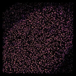
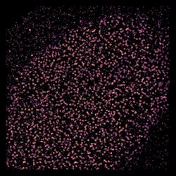
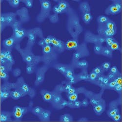
For instance, new super-resolution images have provided the clearest views yet of white blood cells fighting infections and cancer.1 Produced by researchers at the University of Manchester (England), the images show how the surface molecules on the cells reorganize when activated by a type of protein found on viral-infected or tumor cells. Professor Daniel Davis, who has led the immune cells investigation, said the team was surprised to learn that immune cell surfaces alter at the nanoscale, and notes that this fact may change the cells' ability to respond in subsequent encounters with diseased tissue. "We have shown that immune cell proteins are not evenly distributed as once thought, but instead they are grouped in very small clumps," he explained.
The work could provide important clues for disease treatment. "Looking at our cells in this much detail gives us a greater understanding about how the immune system works and could provide useful clues for developing drugs to target disease in the future."
Along similar lines, scientists at the European Molecular Biology Laboratory (EMBL; Heidelberg, Germany) have used stochastic super-resolution microscopy to solve a decade-long cell-biology debate.2 The debate centered on the structure of the nuclear pore complex, which controls access to the genome by acting as a gate into a cell's nucleus. Electron tomography studies had previously shown the gate's overall shape. And such techniques as x-ray crystallography and single-particle electron microscopy had shown that the ring around the nucleus' wall is formed by 16 or 32 copies of Y-shaped building blocks formed by nine proteins each. But researchers didn't know how the Ys are arranged to form a ring.
The EMBL scientists labeled a series of points along each arm and tail of each Y using fluorescent tags, and then imaged them with a stochastic super-resolution microscope. By combining images from thousands of nuclear pores, and by using single-particle averaging, they determined the average positions of fluorescent molecular labels with a precision well below 1 nm. The result was a rainbow of rings whose order and spacing meant the Y-shaped molecules in the nuclear pore must lie in an orderly circle around the opening, all with the same arm of the Y pointing toward the pore's center. "When we looked at our images, there was no question: they have to be lying head-to-tail around the hole," said lead author Anna Szymborska.
Now the scientists intend to delve deeper into the mysteries of the nuclear pore—to determine whether the circle of Ys is arranged clockwise, studying it at different stages of assembly, looking at other parts of the pore, and investigating it in three dimensions. They will also begin trying to understand the "machines" behind cell division, which are similarly built from hundreds of pieces.
1. S. V. Pageon et al., Sci. Signal., 6, ra62 (2013).
2. A. Szymborska et al., Science, 341, 6146, 655–658 (2013); doi:10.1126/science.1240672.


