ADVANCED MICROSCOPY/SUPERCONTINUUM WHITE LIGHT: Multiple microscopy modes in a single sweep with supercontinuum white light
ByLioba Kuschel and Rolf T. Borlinghaus
Lasers have been critical to the advancement on confocal microscopy, and the white light laser (WLL) offers particular advantages. Finessing WLL output for bioimaging is a complex task, though, and traditional approaches retain key limitations. But acousto-optical beamsplitting enables smoother operation, leading to enhanced microscopy capabilities.
The invention and development of lasers as light sources was one of the critical events enabling the enormous success of confocal microscopy in biological sciences.1-3 The features of lasers that are key for this application—high luminance and a nearly 100% collimated beam—allow sufficient light to be focused in a diffraction-limited spot, forming the basis of confocal imaging. The temporal coherence of laser beams is not required for this microscopy technique and can even be a nuisance, as it creates interference effects that must be avoided through careful design.
A real limitation of lasers is their monochromy, which requires a whole battery of lasers to be combined in order to perform multi-parameter fluorescence measurements in simultaneous mode, a method expected as a standard by biomedical researchers. This obstacle was cleared with the advent of supercontinuum sources, which provide white emission along with high luminance and laser-type beam profiles.
But supercontinuum sources are not directly applicable to bioimaging because of the delicate nature of specimens. Now, their adaptation has taken a leap forward.
White light lasers and supercontinuum fiber
The white light laser (WLL) takes advantage of a series of nonlinear effects that occur in all optical materials. Commonly, these effects are not seen, as their probability is too low in ordinary optical elements (usually, they are unwanted, too). The supercontinuum fiber3 enhances these effects in order to create a broad spectrum out of a monochromatic source. The heart of the fiber is an array of some-hundred hollow tubes, usually arranged in a hexagonal pattern and also referred to as photonic crystal fibers (PCFs). To generate sufficient white light, a high photon density is required.
To provide this density, a pulsed illumination is introduced. The first stage of a WLL source is therefore a seed laser—a fiber laser that emits infrared (IR) light in a pulsed mode (typically 80 MHz). The second stage is designed as an amplifier, boosting the total output to some watts and still keeping the pulsed nature of the seed source. Consequently, the first two stages provide very high, intense IR pulses that are fed into the supercontinuum fiber.
The nonlinear effects in the PCFs generate a broad spectrum, spanning a couple of hundred nanometers, including the visible range. The output consequently appears as white emission. This white light is not a combination of three colors, unlike pseudo-white sources such as Ar/Kr lasers that provide blue, green, and red monochrome lines—which the brain combines to white. The WLL emits a truly white spectrum, like an incandescent bulb or the sun. But it has the beam properties of a laser, which is required for true confocal microscopy.4
For confocal imaging
The light requirements for confocal imaging are intensity of a few milliwatts (otherwise, the sample would burn away immediately) along with small bandlets of spectral range that can specifically excite the desired fluorochromes. WLL provides an intensity of some watts, so for this application the white emission is typically filtered with a dynamic device, an acousto-optical tunable filter (AOTF)—a crystal that is electronically programmable to separate the desired colors out of the white spectrum. It creates a small spectral bandlet only a few nanometers wide (for instance, 0.5 to 2 nm) and whose color is programmable and thus steplessly tunable: The operator can pick whatever color is needed for the application. The intensity of such a bandlet is in the range of a couple of milliwatts—still too high for standard confocal imaging. To control the intensity, a second parameter is available that allows stepless tuning of the brightness between zero and the maximum.
Thus, the source offers light that is tunable in both color and intensity over the full range of visible light. The appropriate color for any fluorochrome can be dialed and illumination intensity is adjustable to fit the current requirements. As if this were not enough, the AOTF allows a whole series of bandlets to be controlled simultaneously; currently, a standard is eight different colors at a time. Each of these bandlets has the properties described above: Free tunability of both color and intensity independent of each other. This is the ideal light source for multicolor fluorescence confocal microscopy; the long-sought "magic" illumination source.
Boundless spectral freedom
Having such a source, the next question is: How can this tunability be kept active when coupled into an incident-light fluorescence device? Usually, the light in such systems is coupled into the beam path by means of a beamsplitter. For fluorescence applications, dichroic beamsplitters are used to cause a certain color to be reflected and other colors to be transmitted. Modern multiband dichroic beamsplitters even allow a series of different colors to be fed in for excitation and transmit the emission between these excitation bands. Nevertheless, the properties of such beamsplitters are fixed by design, and a switch to a different set of excitation colors consequently requires a switch of the beamsplitter as well. If the light source is continuously tunable with a series of colors simultaneously, a nearly infinite number of dichroic beamsplitters would be required. This is technically impossible. As an alternative, the beamsplitter could be a grey-splitter, but this would reject a significant portion of precious fluorescence photons throughout the whole spectrum.
A design that addresses all these needs is the acousto-optical beamsplitter (AOBS). Like AOTF, this device is based on acousto-optical crystals but it operates in reverse mode. It allows simultaneous injection of a series of spectral bandlets (currently, a standard is eight) for excitation of multiple fluorochromes while being transparent for efficiently collecting the fluorescence emission in between. And in contrast to glass filter-based dichroics, the device is fully tunable. Any combination of excitation colors within the visible range is possible. This flexibility comes with simple operation: When a set of required colors is selected from the light source, the AOBS is automatically set to the appropriate modes. The operator does not need to think about which beamsplitter is required and there is no danger of accidentally using the wrong splitter device.
As the excitation color is steplessly tunable, a new confocal application method becomes available: excitation spectral imaging. Upon setting a range of excitation wavelengths, a series of images with incremented excitation wavelength can be recorded. The AOBS will continuously follow the incrementing excitation and allow perfect excitation and emission performance at any wavelength.
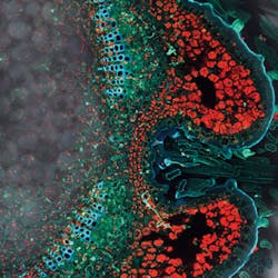
The spectral excitation imaging can be combined with spectral emission imaging as well. With a spectral detection device that allows stepless selection of any emission band, an additional method is available: spectral two-dimensional excitation emission mapping ("Lambda Square" image acquisition). At each excitation color, a full spectrum of the emission is recorded. As this recording is compiled as an image series, the excitation emission properties of selected structures and areas of the sample under examination may be evaluated.
So far, we have discussed the spectral possibilities of a WLL source, and demonstrated the conceivable applications now available with respect to spectral emission and excitation. But because WLL is a pulsed source, another series of very different applications also becomes available.
Fluorescence lifetime imaging and gated detection
During the fluorescence process, molecules are promoted from the ground state to an excited state by absorption of a photon. The excited state rapidly relaxes into the lowest vibrational state, from which energy is then emitted by a photon with less energy than the excitation (Stokes shift). This emission is temporally delayed, typically by a couple of nanoseconds. The average time a molecule stays in that excited state correlates to the chemical compound and is referred to as fluorescence lifetime. With a sufficiently short excitation pulse, the fluorescence will decay exponentially. This decay is measured with time-correlated single photon counting (TCSPC), which ultimately provides a complete decay curve from a given sample. A curve fit on the decay will provide the characteristic decay time(s) and thus information on the fluorescence lifetime of the molecules in the sample.
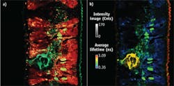
The pulse width of the WLL is below 100 ps and therefore short enough to separate excitation from emission photons in time. The pulse rate is about 80 MHz, corresponding to a pulse distance of 12.5 ns—longer than typical fluorescence lifetimes. The TCSPS method is well established with pulsed single-frequency lasers and IR lasers, often used for multiphoton excitation. The resulting data are images that contain fluorescence lifetime information in each pixel (as compared to intensity information in ordinary imaging). Lifetime measurement is conveniently implemented in systems with WLL sources. In contrast to classical approaches, WLL-based systems allow stepless tuning of the excitation color. Therefore, it becomes possible to measure fluorescence lifetime as a function of excitation wavelength.5
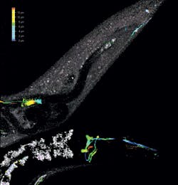
Fluorescence lifetime imaging allows the separation of spectrally identical fluorochromes. Upon illumination with one wavelength and emission collection in a single band, it is possible to see two or three different fluorescence species that differ only in terms of fluorescence lifetime. Of course, the method enables both excitation and emission to be varied, and allows recording of intensity and lifetime dependence in two spectral dimensions. And because confocal was originally invented for spatial three-dimensional data recording, all these acquisition modes can be applied in combination with 3D stacking.
The pulsed excitation allows yet another approach to acquire data in a true confocal microscope system. In combination with a sensor device that is fast enough to allow photon counting at high rates and has sufficient dynamic range to cover the typical intensities in fluorescence imaging, a "pulsed detection" is accessible. Here, the signal is collected only if the collection gates are open; if not, it is suppressed. These modes became available with the introduction of modern hybrid sensor technology. Hybrid detectors (HyD) are composed of classical vacuum and modern silicon-based technologies. Contrary to classic photomultiplier tubes (PMTs), HyDs use a single-step electron acceleration in vacuum, thus at a very high voltage (for example, 8000 V). The (single) photoelectron gains considerable energy during this acceleration and dissipates when it hits a silicon target, generating some 1500 charges. This corresponds to amplification of 1500x. The silicon target is combined with a sequence of silicon layers that make up an avalanche photodiode (APD), generating another 100x amplification. This is sufficient to significantly separate the photon-induced pulses for photon counting, even at comparably high photon rates.
With these devices, it becomes possible to gate detection within the excitation pulses. Data acquisition in a single pixel lasts approximately 100 μs to 30 ns, depending on scan frequency and the number of pixels recorded per line. Within this pixel duration time, the pulsed laser will fire repeatedly—at least two to three times in the very short pixel times with the resonant scanner at high resolution. Usually, all photons would be collected during that time and separated spectrally only by means of filters or true spectral detectors. Photon counting with HyDs allows selection of the events that are to contribute to the final signal and those that are to be omitted. So for example, it is possible to remove all photons that are collected during the excitation pulse time (convoluted with the instrument response time). This will produce an image free of light that is stray or reflected from the excitation source without any spectral filtering. The method is called "gated detection," as the detection is restricted to a gate—in this case, the time between the excitation pulses. This gated detection is independent of colors; it operates in a spectrally "white" mode and does not need to adapt different optical elements when changing excitation and emission colors.
As time gates can be tuned within the pulse repetition time, it is also possible to funnel photons into sequential channels based on how quickly they follow the excitation pulse: Early photons correlate with short lifetimes, whereas late photons correspond to longer lifetimes. When collecting these photons into sequential channels, it is immediately possible to separate fluorescent species that have sufficient differences in fluorescence lifetime—without requiring dedicated fluorescence lifetime imaging microscopy (FLIM) equipment. Fluorescence decay, excitation wavelength, and emission color are thus easily accessible to perform multimodal imaging in a system that is equipped with a WLL and HyDs.
Last but not least, the gated detection upon pulsed excitation with a WLL source also allows improved stimulated emission depletion (STED) super-resolution imaging by restricting data collection of late photons.
WLL offers many advantages, and new technology for adapting it to bioimaging tasks both enhances and broadens its applicability.
References
1. T. H. Maiman, Nature, 187, 4736, 493-494 (1960).
2. C. J. R. Sheppard and A. Choudhury, Opt. Acta, 24, 1051-1073 (1977).
3. P. Russell et al., Science, 299, 358 (2003).
4. R. T. Borlinghaus and L. R. Kuschel, "The White Confocal - Spectral Gaps Closed," Current microscopy contributions to advances in science and technology, Formatex Research Center, Badajoz, Spain (December 2012).
5. R. Borlinghaus, "The White Confocal: Continuous Spectra Tuning in Excitation and Emission," Optical Fluorescence Microscopy - From the Spectral to the Nano Dimension, Springer Berlin Heidelberg, Berlin, Germany (2011).
Lioba Kuschel is product manager and Rolf T. Borlinghaus is scientific advisor with Leica Microsystems CMS (Mannheim, Germany; www.leica-microsystems.com). Contact Dr. Kuschel at [email protected].


