Synchronized laser therapy gets people back on the right foot quickly
ByLEE MATHER
Ed Davis, DPM, FACFAS, a podiatrist based in the San Antonio, TX, area, has boosted his treatment options in his practice with Multiwave Locked System (MLS) therapy for treating foot and ankle pain, inflammation and edema, and for repairing superficial lesions.
MLS therapy combines a continuous/chopped 808 nm emission with a 905 nm pulsed emission to achieve an intense, anti-edema and anti-inflammatory effect and a strong, analgesic effect, respectively. The patented control system that generates the MLS pulse synchronizes the two emissions, allowing the anti-inflammatory and anti-edema effects to reinforce each other. Davis uses the MIX5 system manufactured by ASA S.r.l. (www.asa-laser.com/uk-2.html; Arcugnano, Italy) in his practice, which received FDA clearance in September 2005.
On average, patients receive a series of three to five treatments spaced one to three days apart; however, there is no limit to the number of treatments, Davis told BioOptics World. A patient with an acute problem due to a non-recurring injury often experiences fairly rapid, lasting results, while someone with a non-recurring injury may require more frequent treatment, coupled with attention to the biomechanical etiology of the injury, he notes.
MLS therapy's synchronized emissions allow for pain to disappear four to 12 hours after treatment if the root cause of the problem is addressed, notes Davis.
For additional information, visit www.southtexaspodiatrist.com.
Award-winning fluorescence image explores the spread of malaria
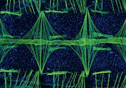
A fluorescence image that takes viewers inside the heart of a mosquito is this year's winner in Nikon's Small World 2010 photomicrography competition (www.nikonsmallworld.com). Vanderbilt University (Nashville, TN) graduate student Jonas King, a member of the research group of Julián Hillyer, took the 100X magnification image while conducting research on the circulatory system of Anopheles gambiae, a mosquito that spreads malaria.
In showing the mosquito heart's structure, King used green fluorescent dye to bind with muscle cells and show the underlying musculature, and blue fluorescent dye to bind with cellular DNA and show the presence of all the cells. The mosquito's body is shown horizontally with its head to the left, and the narrow tube that runs horizontally across the middle is the heart. The muscles that wind around the heart show up clearly in green.
Hillyer notes that because this particular mosquito's circulatory system plays a crucial part in spreading the malaria parasite, their research group is working to better understand its biology to develop pest- and disease-control strategies.
More BioOptics World Current Issue Articles
More BioOptics World Archives Issue Articles


