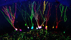
Fluorescent cell marking could lead to deeper understanding of brain function
In neuroscience, it is a challenge to individually label cells and to track them over space or time. Our brain has billions of cells and to be able to distinguish them at the single-cell level, and to modify their activity, is crucial to understand such a complex organ. Knowing this, scientists at the University of Southampton in England have developed a fluorescent technique to mark individual brain cells to help improve our understanding of how the brain works.
The new marking technique, known as multicolor red-green-blue (RGB) tracking, allows single cells to be encoded with a heritable color mark generated by a random combination of the three basic colors.
Brains are injected with a solution containing three viral vectors, each producing one fluorescent protein in each of the three colors. Each individual cell will take on a combination of the three colors to acquire a characteristic watermark. This approach allows researchers to color-code cells that would otherwise not be visible and undistinguishable from each other.
Once the cell has been marked, the mark integrates into the DNA and will be expressed forever in that cell, as well as in any daughter cells.
Dr. Diego Gomez-Nicola, a Career Track Lecturer and MRC NIRG Fellow in the Centre for Biological Sciences at the University of Southampton, who led the multicolor RGB tracking research, says: "With this technique, we have proved the effective spatial and temporal tracking of neural cells, as well as the analysis of cell progeny. This innovative approach is primarily focused to improve neuroscience research, from allowing analysis of clonality to the completion of effective live imaging at the single-cell level.
"We predict that the use of multicolor RGB tracking will have an impact on how neuroscientists around the world design their experiments. It will allow them to answer questions they were unable to tackle before and contribute to the progress of understanding how our brain works."
For the researchers, the next step is to change the physiology or identity of certain cells by driving multiple genetic modification of genes of interest with the RGB vectors. In the same way they made cells express fluorescent proteins, researchers hope they can change the cell expression of target genes, which would underpin gene therapy-based therapeutic approaches.
Full details of the research appear in the journal Scientific Reports; for more information, please visit http://dx.doi.org/10.1038/srep07520.
-----
Follow us on Twitter, 'like' us on Facebook, connect with us on Google+, and join our group on LinkedIn
Subscribe now to BioOptics World magazine; it's free!

