Super-resolution microscopy technique makes key DNA finding for possible disease treatments
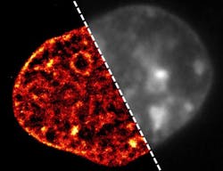
Using a super-resolution microscopy technique, scientists at the Institute of Molecular Biology (IMB) at Johannes Gutenberg University Mainz (JGU; Mainz, Germany) have been able to see the dramatic changes that occur in the DNA of cells that are starved of oxygen and nutrients. This starved state is typical in some of today's most common diseases, particularly heart attack, stroke, and cancer. The findings provide new insight into the damage these diseases cause and may help researchers to discover new ways of treating them.
Related: Super-resolution microscopy helps quantify viral DNA
When a person has a heart attack or a stroke, the blood supply to part of their heart or brain is blocked. This deprives affected cells there of oxygen and nutrients, a condition known as ischemia, and can cause long-term damage, meaning that the person may never fully recover. Ina Kirmes, a PhD student in the group of Dr. George Reid at IMB, investigated what happens to the DNA in cells that are cut off from their oxygen and nutrient supply.
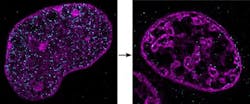
In a healthy cell, large parts of the DNA are open and accessible. This means that genes can be easily read and translated into proteins, so that the cell can function normally. However, the researchers showed that in ischemia, DNA changes dramatically: it compacts into tight clusters. The genes in this clumped DNA cannot be read as easily anymore by the cell, their activity is substantially reduced, and the cell effectively shuts down. If cells in a person's heart stop working properly, this part of the heart stops beating and they will have a heart attack. Similarly, when blood supply is blocked to cells in someone's brain and their cells there are starved of oxygen and nutrients, they have a stroke. Reid says that with this finding, they can start to look at ways of preventing this compaction of DNA.
Key to the discovery was a close collaboration with Aleksander Szczurek, joint first author on a paper describing the work, who is part of the group of Professor Christoph Cremer at IMB. They developed a new method that made it possible to see DNA inside the cell at a level of detail never achieved before. Their technique is a further development of super-resolution microscopy that they call single-molecule localization microscopy (SMLM), which uses blinking dyes that bind to DNA to enable the researchers to define the location of individual molecules in cells.
Full details of the work appear in the journal Experimental Cell Research; for more information, please visit http://dx.doi.org/10.1016/j.yexcr.2015.08.020.
Follow us on Twitter, 'like' us on Facebook, connect with us on Google+, and join our group on LinkedIn

