Fluorescence microscopy add-on shows promise for more precise neurosurgery
A team of researchers at the University of Arizona (UA; Tucson, AZ) has developed a fluorescence microscopy device that allows neurosurgeons to see blood flowing inside vessels and more clearly distinguish cancerous from healthy tissue under the microscope.
Related: Advanced surgery: NIR fluorescence guidance arrives
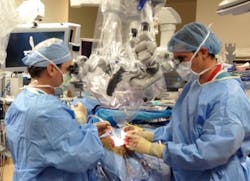
Called augmented microscopy, the technology gives surgeons a much more detailed picture in real time and helps them stay on course in surgeries where being off 2 mm could cause paralysis, blindness, and even death. And surgeons get this better view without having to learn new technical skills or adapt to changes in the operating room.
The new technology overlays a brightfield image that a surgeon sees under a microscope with an electronically processed image using near-infrared (NIR) fluorescence, a computer-generated imaging technology in which contrast agents are injected in patients to illuminate vital diagnostic information and help surgeons avoid cutting the wrong vessel or removing healthy tissue.
Most neurosurgeons must look up from a surgical microscope (stereomicroscope) to view fluorescence on a display monitor. If they have a microscope adapted to project fluorescence, it switches back and forth between the real and electronic views, the surgeons' field of vision momentarily fading to black in between. Further, the fluorescence shows only contrast in black and white, not anatomical structures or their spatial relationships. So surgeons must visualize how fluorescence lines up with the anatomical structures they see under the microscope. The newly developed add-on removes such interruptions or guesswork by showing surgeons real and fluorescence images simultaneously and in one location.
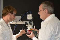
The new device, a small box fitted inside a surgical microscope, combines electronic circuitry and optical technologies to superimpose the fluorescence image on the real one and send the augmented view up through the microscope's right eyepiece to the surgeon.
Perhaps the most valuable application for augmented microscopy is treating brain cancer, says Marek Romanowski, a UA associate professor of biomedical engineering who holds appointments with the University of Arizona Cancer Center and BIO5 Institute. Current surgical microscopes limit how much of the cancer tissue surgeons can see and how precisely they can determine its boundaries, he says.
Full details of the work appear in the Journal of Biomedical Optics; for more information, please visit http://dx.doi.org/10.1117/1.JBO.20.10.106002.
Follow us on Twitter, 'like' us on Facebook, connect with us on Google+, and join our group on LinkedIn
BioOptics World Editors
We edited the content of this article, which was contributed by outside sources, to fit our style and substance requirements. (Editor’s Note: BioOptics World has folded as a brand and is now part of Laser Focus World, effective in 2022.)

