Optical tomography technique enables diagnostic 3D image capture in real time

Feb. 9, 2015
A technique developed by researchers in Spain that uses optical projection tomography makes it possible to use optical biomarkers often used with transgenic animals, such as green fluorescent protein (GFP), to follow the development of living organisms up to 3 mm long with three-dimensional (3D) images.

A technique developed by researchers at Charles III University of Madrid (UC3M) in Spain that uses optical projection tomography makes it possible to use optical biomarkers often used with transgenic animals, such as green fluorescent protein (GFP), to follow the development of living organisms up to 3 mm long with three-dimensional (3D) images.

Related: Biomedical diagnostics using personalized 3D imaging possible

Researcher Jorge Ripoll of the UC3M Department of Bioengineering and Aerospace Engineering, who was involved in the work, says that the advance consists of being able to follow the development of these organisms, which normally appear opaque when viewed with a conventional microscope because they diffuse a lot of light when they approach adulthood.

"It helps us visualize new stages," says Ripoll. He adds that although the method cannot be used on living humans because their tissue is very opaque, it can be used to take 3D measurements of biopsies, which is valuable to surgeons.

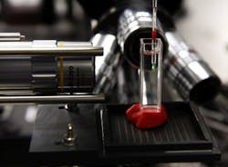
A new technique that uses optical projection tomography can capture real-time diagnostic 3D images.

Ripoll explains that their system consists of a light source that stimulates the fluorescence and a camera to detect it, and requires that the sample rotates much like how x-rays are taken. Afterwards, with that information, "we must construct a three-dimensional image," he explains.

Full details of the work appear in the journal Scientific Reports; for more information, please visit http://dx.doi.org/10.1038/srep07325.

------

Follow us on Twitter, 'like' us on Facebook, connect with us on Google+, and join our group on LinkedIn

Subscribe now to BioOptics World magazine; it's free!
About the Author

BioOptics World Editors

We edited the content of this article, which was contributed by outside sources, to fit our style and substance requirements. (Editor’s Note: BioOptics World has folded as a brand and is now part of Laser Focus World, effective in 2022.)

Sponsored Recommendations

How to Tune Servo Systems: Force Control

Oct. 23, 2024
Tuning the servo system to meet or exceed the performance specification can be a troubling task, join our webinar to learn to optimize performance.

Laser Machining: Dynamic Error Reduction via Galvo Compensation

Oct. 23, 2024
A common misconception is that high throughput implies higher speeds, but the real factor that impacts throughput is higher accelerations. Read more here!

Boost Productivity and Process Quality in High-Performance Laser Processing

Oct. 23, 2024
Read a discussion about developments in high-dynamic laser processing that improve process throughput and part quality.

Precision Automation Technologies that Minimize Laser Cut Hypotube Manufacturing Risk

Oct. 23, 2024
In this webinar, you will discover the precision automation technologies essential for manufacturing high-quality laser-cut hypotubes. Learn key processes, techniques, and best...

Voice your opinion!

To join the conversation, and become an exclusive member of Laser Focus World, create an account today!