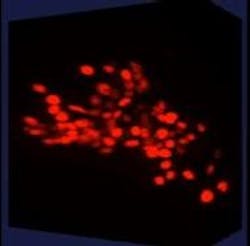
Researchers capture neural activity of nematode brains in 3D
Researchers at Princeton University (New Jersey) have captured recordings of neural activity in nearly the entire brain of a free-moving animal. The 3D recordings could provide scientists with a better understanding of how neurons coordinate action and perception in animals.
Related: New fluorescent protein views neural activity beyond microscope's field of view
The technique the research team used allowed them to record 3D footage of neural activity in the nematode Caenorhabditis elegans (C. elegans), a 1-mm-long worm species with a nervous system containing 302 neurons. The researchers correlated the activity of 77 neurons from the animal's nervous system with specific behaviors, such as backward or forward motion and turning.
Previous work related to neuron activity either focuses on small subregions of the brain or is based on observations of organisms that are unconscious or somehow limited in mobility, explains Andrew Leifer, an associate research scholar in Princeton's Lewis-Sigler Institute for Integrative Genomics and a corresponding author of the study.
A current focus in neuroscience is understanding how networks of neurons coordinate to produce behavior, Leifer says. The technology to record from numerous neurons as an animal goes about its normal activities, however, has been slow to develop, he says. Neural networks are infinitesimal arrangements of chemical signals and electrical impulses that can include, as in humans, billions of cells.
The simpler nervous system of C. elegans provided the researchers with a more manageable testing ground for their instrument. Yet, it also could reveal information about how neurons work together that applies to more complex organisms, Leifer says. For instance, the researchers were surprised by the number of neurons involved in the seemingly simple act of turning around.
The researchers designed an instrument that captures calcium levels in brain cells as they communicate with one another. The level of calcium in each brain cell tells the researchers how active that cell is in its communication with other cells in the nervous system. The researchers induced the nemotodes' brain cells to generate a protein known as a calcium indicator that becomes fluorescent when it comes in contact with calcium.
The researchers used a special type of microscope to record in 3D both the nematodes' free movements and neuron-level calcium activity for more than 4 min. Three-dimensional software designed by the researchers monitored the position of an animal's head in real time as a motorized platform automatically adjusted to keep the animal within the field of view of a series of cameras.
The entire setup drew from various disciplines and techniques, including physics, computer science, and engineering, Leifer says. For instance, the real-time computer vision algorithms the researchers used to track the worms' brains are similar in principle to the ones used in robotics or in self-driving cars.
Even more about the inner workings of the C. elegans nervous system remains to be extracted from the researchers' data over the next year, Leifer says. The team is currently working to flesh out the correlations between neural activity and behavior in general.
"An exciting next step is to use correlations in our recordings to build mathematical and computer models of how the brain functions," Leifer says. "We can use these models to generate hypotheses about how neural activity generates behavior. We plan to then test these hypotheses, for example, by stimulating specific neurons in an organism and observing the resulting behavior."
Full details of the work appear in the Proceedings of the National Academy of Sciences; for more information, please visit http://dx.doi.org/10.1073/pnas.1507110112.
Follow us on Twitter, 'like' us on Facebook, connect with us on Google+, and join our group on LinkedIn
