Bioimaging for applied cell research center to open in Munich
Leica Microsystems (Wetzlar, Germany) and the Biomedical Center (BMC) of Ludwig-Maximilians University (LMU; Munich, Germany) will inaugurate the Leica Bioimaging Center on February 17, 2016. Leica will use the new facility, which is located at the BMC and the result of a strategic collaboration between the company and LMU, as a reference and demo center for light microscopy. The center provides the opportunity for a close cooperation between microscope developers and users to develop innovations in modern light microscopy and establish their application in applied cell research.
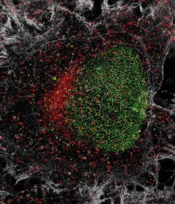
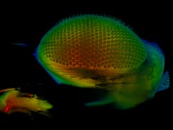
For BMC researchers, the Leica Bioimaging Center offers access to the latest technologies in light microscopy, enabling them to make the smallest structures of the cell visible, even at the nanoscale, and investigate biological processes on the molecular level. Among these technologies are super-resolution stimulated emission depletion (STED) microscopy, which was awarded the Nobel Prize in Chemistry in 2014, and light-sheet microscopy, which has utility with large, living samples.
The Leica Bioimaging Center is the biggest reference and demo center to be established by Leica Microsystems in Europe so far. There, customers and potential customers can discover the company's latest systems, see them used in research onsite, and exchange experiences with the users at the facility. In addition, new products can be tested thoroughly by the users of the core facility before being introduced to the market. The facility will be operated by the Walter-Brendel-Centre of Experimental Medicine, one of the eight resident professorships at BMC.The inauguration of the Leica Bioimaging Center will be accompanied by a scientific symposium on February 17th. The topic "Plasticity of Cell Programs as Visualized by Advanced Microscopy Techniques" is relevant to scientists from various areas, such as physiology, molecular biology, immunology, and cell biology. Participation at the symposium is free, but requests registration at www.leica-microsystems.com/inauguration-symposium.

