Microscopy, molecular methods look at how blood vessels stay healthy
Anna Randi, MD, PhD, and Graeme Birdsey, BSc, PhD, MA, of the National Heart and Lung Institute at the Imperial Centre for Translational and Experimental Medicine (ICTEM; London, England) are working to understand how blood vessels form and repair themselves and the role they play in various diseases, such as heart disease, cancer, and dementia. Combining microscopy and molecular techniques, including fluorescent tagging and gene targeting, are key to their work, as they helped them to identify and track important signaling molecules.
Related: OCT images blood vessels underneath the skin that feed cancer
Endothelial cells, which line the entire circulatory system and control what goes in and what goes out, have been identified as the key to maintaining healthy, happy, and functioning blood vessels, Randi explains. "They form a single sheet, and they do this through cell-to cell-signaling; we still donât know how they do it exactly, but itâs a beautiful mechanism," she says.
In cancer, this process of forming new blood vessels, which is essential for life, goes into overdrive as new blood vessels are sprouted to support the growing tumor. A whole class of new drugs is currently being developed that inhibits angiogenesis (blood vessel formation) and stymies the sustaining blood flow to the tumor, killing it off.
By contrast, in the case of cardiovascular disease, endothelial cells become dysfunctional because of smoking, high blood pressure, and elevated levels of low-density lipoprotein (LDL) cholesterol. Eventually, this can lead to the formation of atherosclerotic plaques, which can narrow the arteries with potentially serious consequences such as myocardial infarction and chronic heart disease. So scientists are trying various ways of stimulating the formation of new vessels in place of the diseased onesa strategy called therapeutic angiogenesis. This is already being tested on patients around the world.
Annaâs research group at Imperial works with cell cultures obtained from the blood vessels of human umbilical cords. Like their native counterparts, these endothelial cells form a single layer on a plastic dish.
One protein that has emerged as particularly interesting is Erg, which Birdsey describes as a 'master regulator' of endothelial cell function. It belongs to a class of protein known as transcription factors, whose job it is to bind to sections of DNA, switching genes on and off and creating a mosaic of genetic expression that programmes the unique identity and function of endothelial cells.
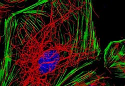
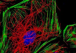
When the research team blocked the action of Erg using so-called âantisenseâ molecules, the contacts between cells destabilized, causing them to leak, Birdsey says. This is because Erg maintains the cellsâ internal scaffolding, made up of actin fibers and a dense web of microtubules.
Further experiments have shown that Erg also has a critical function in endothelial cell migration, which is important for making new vessels as well as repairing them. In one demonstration, Birdsey prepared two separate dishes of endothelial cellsâone with the Erg intact and the other with Erg blocked. He made a cut straight down the middle of the cell sheet and watched them move using time-lapse microscopy. In the Erg-positive dish, the cells at the edge of the simulated wound migrated inward, eventually sealing the gap. But in the Erg-minus dish the cells simply dithered, seemingly not able to coordinate an effective response.
Genetic profiling of endothelial cells has shown that Erg controls a complex web of more than 2,000 genes, themselves involved in migration, angiogenesis, and endothelial cell function. The challenge will be to untangle this web and map the precise relationship between molecules to identify some promising targets for treatment. This could, for example, limit new blood vessel growth in cancer tissue, or promote the formation of new vessels in the heart after myocardial infarction.
For more information, please visit http://www.jbc.org/content/287/15/12331.
-----
Follow us on Twitter, 'like' us on Facebook, and join our group on LinkedIn
Subscribe now to BioOptics World magazine; it's free!

