Fluorescence microscopy method reveals dynamics of telomere DNA structure
Researchers at the University of California, Santa Cruz used a fluorescence microscopy method to reveal structural and mechanical properties of telomeres (the protective caps on the ends of chromosomes). The discovery could help guide the development of new anti-cancer drugs.
Telomeres are long, repetitive DNA sequences at the ends of chromosomes that serve a protective function: as cells divide, their telomeres get progressively shorter until eventually the cells stop dividing. Telomeres can grow longer, however, through the action of an enzyme called telomerase, which is especially active in cells that need to keep dividing indefinitely, such as stem cells. Researchers have also found that most tumor cells show high telomerase activity.
Michael Stone, an assistant professor of chemistry and biochemistry at UC Santa Cruz, says his lab is particularly interested in the folding and unfolding of a DNA structure at the tail end of the telomere, known as a G-quadruplex, because it plays a key role in regulating telomerase activity. "Most cancer cells use telomerase as one mechanism to maintain uncontrolled growth, so it is an important target for anti-cancer therapeutics," he explains. "The G-quadruplex structures of telomere DNA inhibit the function of the telomerase enzyme, so we wanted to understand the mechanical stability of this structure."
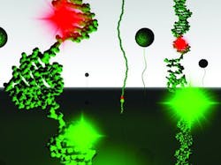
Xi Long, a graduate student in Stone's lab, led the project, which involved integrating two techniques to manipulate and monitor single DNA molecules during the unfolding of the G-quadruplex structure. A "magnetic tweezers" system was used to stretch the DNA molecule, while a fluorescence microscopy method was used to monitor small-scale structural changes in the DNA. The results showed that a relatively small structural displacement causes the G-quadruplex to unfold.
"Unlike other DNA structures, the G-quadruplex structure is fairly brittle. It takes very little perturbation to make the whole thing fall apart," Stone says. "We also found that the unfolded state has a highly compacted conformation, which tells us that it still has interactions that favor the folding reaction."
These findings have implications for understanding the molecular mechanisms of telomere-associated proteins and enzymes involved in the unfolding reaction, as well as for rational design of anti-cancer drugs, Stone says. Small molecules that bind to and stabilize telomere DNA G-quadruplexes have shown promise as anti-cancer drugs.
The integration of fluorescence measurements and magnetic tweezers is a powerful method for monitoring DNA structural dynamics, and as biophysical techniques go, it is not hard to implement, Stone says. His lab worked with DNA molecules containing the G-quadruplex sequence from human telomere DNA, attaching one end of the DNA to a glass slide and the other end to a tiny magnetic bead. A magnet held above the sample pulled on the bead, exerting a stretching force on the DNA molecule that varied according to how close the magnet was to the sample.
At the same time, the researchers used a fluorescence technique called single-molecule Förster resonance energy transfer (FRET) to monitor small-scale structural changes in the DNA. As energy from one fluorescent dye molecule is transferred to a second dye molecule, the efficiency of the energy transfer can be measured in real time. The dye molecules can be coupled directly to the DNA molecule at specific sites, allowing researchers to monitor the molecular dynamics of the system as it is being manipulated by the magnetic tweezers.
Full details of the work Nucleic Acids Research; for more information, please visit http://nar.oxfordjournals.org/content/early/2013/01/08/nar.gks1341.full.
-----
Follow us on Twitter, 'like' us on Facebook, and join our group on LinkedIn
Subscribe now to BioOptics World magazine; it's free!
