Sugar solution makes tissues see-through, enables images with unprecedented detail
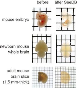
Researchers from the RIKEN Center for Developmental Biology (Kobe, Japan) have developed a new sugar- and water-based solution that turns tissues transparent in three days without disrupting the shape and chemical nature of the samples. Combined with fluorescence microscopy, this technique enabled them to obtain detailed images of a mouse brain at an unprecedented resolution.
Related: Microscopy method renders spinal cord tissue transparent
Over the past few years, teams in the U.S. and Japan have reported a number of techniques to make biological samples transparent that have enabled researchers to look deep down into biological structures like the brain.
âHowever, these clearing techniques have limitations because they induce chemical and morphological damage to the sample and require time-consuming procedures,â explains Dr. Takeshi Imai, who led the study.
SeeDB, an aqueous fructose solution that Dr. Imai developed with colleagues Drs. Meng-Tsen Ke and Satoshi Fujimoto, overcomes these limitations. Using SeeDB, the researchers were able to make mouse embryos and brains transparent in just three days without damaging the fine structures of the samples or the fluorescent dyes they had injected in them.
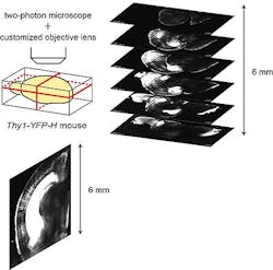
They could then visualize the neuronal circuitry inside a mouse brain, at the whole-brain scale, under a customized fluorescence microscope without making mechanical sections through the brain. In their study, they also describe the detailed wiring patterns of commissural fibers connecting the right and left hemispheres of the cerebral cortex, in three dimensions, for the first time.
Dr. Imai and colleagues report that they were also able to visualize in three dimensions the wiring of mitral cells in the olfactory bulb, which is involved the detection of smells, at single-fiber resolution.
âBecause SeeDB is inexpensive, quick, easy and safe to use, and requires no special equipment, it will prove useful for a broad range of studies, including the study of neuronal circuits in human samples,â explain the authors.
Full details appear in Nature Neuroscience; for more information, please visit http://www.nature.com/neuro/journal/vaop/ncurrent/full/nn.3447.html.
-----
Follow us on Twitter, 'like' us on Facebook, and join our group on LinkedIn
Subscribe now to BioOptics World magazine; it's free!

