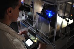
New microscopy lab promising for advancing imaging methods
The Neurobiology Centre of the Nencki Institute of Experimental Biology (Warsaw, Poland) has opened a new laboratory that combines light and electron microscopy methods in efforts to advance microscopic imaging. Called the Laboratory of Imaging Tissue Structure and Function and established as part of the Centre for Preclinical Research and Technology (CePT), the new lab is a core research facility that will focus on three-dimensional mapping of the internal structure of nervous cells and microscopic brain observation in living organisms.
Related: Light-absorbing engineered protein improves usefulness of electron microscopy for bioimaging
Tytus BernaÅ, Ph.D., who heads the new lab, explains that their combination approach will help the researchers obtain more comprehensive images of cell and tissue physiology and more precise images of their structure. What's more, he says, they will be able collect information at the microscopic level about what is going on in live nervous tissue.
Research being carried out in the new lab is using confocal and two-photon microscopy, time-resolved imaging, and super-resolution and correlative microscopy. What's especially interesting is the fluorescence confocal microscope operating in tandem with the electron microscope. This setâwhich is unique in Polandâcombines high-resolution, characteristic for electron imaging techniques, with the wealth of biological information provided by images obtained by using light. An additional advantage of this set is its capability for automatic imaging of a 3D structure of tissue and cells preparations. The resolution of these devices is so great that it is possible to observe not only whole cells, but also the internal structure of their parts (such as only cell nuclei or axons).
Since neurobiological research dominates the scientific activity of the Nencki Institute, an important piece of equipment in the new lab is a microscope for quick imaging of thick fragments of live nervous tissue. It allows researchers microscopic observation of changes occurring in the brains of mice and ratsâpractically in real time. These observations will help study brain plasticity, which plays an important role in mental processes related to learning, memory, cognitive aging, Alzheimerâs and Parkinsonâs diseases, or regaining mental fitness following a stroke.
In the coming months, the lab's researchers will have completed building their own super-resolution microscope that uses adaptive optics to model wavefront of light, explains BernaÅ. This device, constructed in cooperation with the team of Maciej Wojtkowski, MSc, Ph.D., from the Faculty of Physics of the Nicolaus Copernicus University (ToruÅ, Poland), will use the sample itselfâwhich usually hinders microscopic imagingâas an additional optical element. As a result, scientists expect to achieve higher resolution than that enabled by the microscope's optics. To analyze the images, they will use algorithms developed by BÅażej Ruszczycki, Ph.D., from the Nencki Institute.
CePT, of which the Nencki Institute is a partner, is the largest biomedical and biotechnological undertaking in Central and Eastern Europe. The budget of this project amounts to over $118.6 million, of which 85% comes from the European Fund for Regional Development. Under CePT, a network of related core facility labs is being established, integrating research and implementation activities of various scientific institutions of the Ochota Research Centre (also in Warsaw). These labs conduct basic and preclinical research at the highest European level in the area of protein structural and functional analysis, physics, chemistry and nanotechnology of biomaterials, molecular biotechnology, instrumental support for medical technologies, pathophysiology and physiology, oncology, genomics, neurobiology, and aging-related diseases.
-----
Follow us on Twitter, 'like' us on Facebook, and join our group on LinkedIn
Subscribe now to BioOptics World magazine; it's free!
