Super-resolution microscopy of large fields in living cells possible with sCMOS

Oct. 31, 2014
Super-resolution microscopy using a novel fluorescence microscope has enabled a team of researchers to perform real-time nanoscopic imaging of large fields of living cells for the first time.

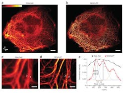
Super-resolution microscopy using a novel fluorescence microscope has enabled researchers from the Max Planck Institute (Göttingen, Germany) to perform real-time nanoscopic imaging of large fields of living cells for the first time. The microscope, which is equipped with a low-noise, high-speed 5.5 Mpixel scientific CMOS (sCMOS) camera, delivers massive parallelization techniques to create 116,000 simultaneous scanning points and super-resolve 120 × 100 µm fields in <1 s.

Related: Nobel Prize honors super-resolution optical microscopy

The research was led by 2014 Nobel Prize winner Prof. Stefan Hell, who first advanced stimulated emission depletion (STED) and a technique dubbed Reversible Saturable/Switchable Optical Fluorescence Transitions (RESOLFT), both of which are far-field, super-resolution microscopy methods. Although RESOLFT can capture images at video rates, imaging speed has been governed by the kinetics of fluorophore state transition and, more importantly, the number of scanning steps required to cover the field of view—until now.

Live-cell imaging with parallelized RESOLFT nanoscopy. The 120 × 100 µm field of view (wide field [a] and RESOLFT [b]) shows PtK2 cells expressing keratin 19–rsEGFP(N205S). The RESOLFT image was reconstructed from 144 frames, each acquired in 22 ms; total image acquisition time was ~3 s. Scale bars = 10 µm. Intensity is from black, low, to white, high. A magnified region (wide field [c] and RESOLFT [d]) of a PtK2 cell expressing keratin 19–rsEGFP(N205S) is represented in (c) and (d), while normalized intensity profiles (e) of the regions between the arrowheads in (c) and (d) are shown with black squares and red dots, respectively. The profile of the RESOLFT data (red line) is fitted to a sum of three Gaussians (purple, green, and orange lines) with individual full-width half-maximum (FWHM) of 77 nm, 133 nm, and 110 nm.

Hell and his research team reconciled the major goals of nanoscopy development: low-intensity operation, large fields of view, and fast recording, at a resolution not limited by diffraction. They demonstrated that RESOLFT nonlinear structured illumination can be parallelized using two incoherently superimposed orthogonal standing light waves. The intensity minima of the resulting pattern act as imaging "doughnuts," providing isotropic resolution in the focal plane and making pattern rotation redundant.

Full details of the work appear in the journal Nature Methods; for more information, please visit http://dx.doi.org/10.1038/nmeth.2556.

-----

Follow us on Twitter, 'like' us on Facebook, connect with us on Google+, and join our group on LinkedIn

Subscribe now to BioOptics World magazine; it's free!
Sponsored Recommendations

Advancing Neuroscience Using High-Precision 3D Printing

March 7, 2025
Learn how Cold Spring Harbor Laboratory Used High-Precision 3D Printing to Advance Neuroscience Research using 3D Printed Optical Drives.

From Prototyping to Production: How High-Precision 3D Printing is Reinventing Electronics Manufacturing

March 7, 2025
Learn how micro 3D printing is enabling miniaturization. As products get smaller the challenge to manufacture small parts increases.

Sputtered Thin-film Coatings

Feb. 27, 2025
Optical thin-film coatings can be deposited by a variety of methods. Learn about 2 traditional methods and a deposition process called sputtering.

What are Notch Filters?

Feb. 27, 2025
Notch filters are ideal for applications that require nearly complete rejection of a laser line while passing as much non-laser light as possible.

Voice your opinion!

To join the conversation, and become an exclusive member of Laser Focus World, create an account today!