Two-photon microscopy method images neuronal activity across the entire brain
Although the entire nervous system connectivity map of the nematode model organism, Caenorhabditis elegans (C. elegans), has been known for more than 25 years, it has not been possible to predict all functional connections underlying behavior. Now, the Vaziri and Zimmer research groups at the Institute of Molecular Pathology (IMP; Vienna, Austria) have demonstrated a two-photon microscopy method that allows neuronal activity across the entire brain to be recorded simultaneously.
Related: High-resolution serial two-photon tomography images whole brains fast
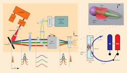
The research groups, partially led by Prof. Alipasha Vaziri, turned to a 5.5 Mpixel scientific CMOS (sCMOS) camera to image the entire volume associated with the worm's brain (75 × 75 × 30 μm) 4–6 times/s using wide-field temporal focusing (WF-TeFo) two-photon microscopy. A novel calcium-indicator enabled them to visualize the activity of a neuron by increasing its fluorescence and permitted unambiguous discrimination of individual neurons within the densely packed head ganglia, explains Vaziri.
WF-TeFo two-photon microscopy allows users to independently control the axial and lateral confinement of the excitation area while taking advantage of the high depth penetration and low scattering properties of two-photon excitation. For their volumetric imaging, the research group scanned an excitation area measuring approx. 60 μm in diameter with an axial confinement measuring approx. 1.9 μm in the axial direction only to speed up the volume acquisition time. A volume acquisition rate approaching 13 Hz was achievable but imaging was performed at speeds of 4–6 Hz, corresponding to approx. 5.6 Mvoxels/s, achieving a lateral spatial resolution of 0.285 μm.
What's more, the method can also be applied straightforwardly to other organisms or more general imaging tasks, and since their novel calcium reporter is a very powerful tool that can be used by all neuroscientists working on C. elegans, their technique has already had a large impact, explains Orla Hanrahan, application specialist – life sciences at Andor Technology (Belfast, Northern Ireland). "If a lab-on-a-chip device is deployed for stimulus delivery in the future, this method provides an enabling platform for establishing functional maps of neuronal networks," she adds.
The work appears in the journal Nature Methods; for more information, please visit http://www.nature.com/nmeth/journal/v10/n10/full/nmeth.2637.html.
