New cyan fluorescent protein yields more sensitive cell imaging
Scientists at the Institut de Biologie Structurale/CNRS-CEA-Université Joseph Fourier (Grenoble, France) have developed a molecule that emits turquoise light more efficiently than ever seen before in living cells, improving the sensitivity of cellular imaging. The teamâled by Antoine Royantâalso comprised scientists from the University of Amsterdam in The Netherlands, the University of Oxford in England, and the European Synchrotron Radiation Facility (ESRF) in Grenoble.
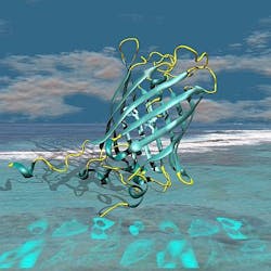
First, using highly brilliant X-ray beams at the ESRF, the scientists from Grenoble and Oxford uncovered subtle details of how cyan fluorescent proteins (CFPs) store incoming energy and retransmit it as fluorescent light. They produced tiny crystals of many different improved CFPs and resolved their molecular structures. These structures revealed a subtle process near the so-called chromophore, the light-emitting complex inside the CFPs, whose fluorescence efficiency could be modulated by the environment. "We could understand the function of individual atoms within CFPs and pinpoint the part of the molecule that needed to be modified to increase the fluorescence yield," says David von Stetten from the ESRF.
In parallel to this work, the Amsterdam team, led by Theodorus Gadella, used an innovative screening technique to study hundreds of modified CFP molecules, measuring their fluorescence lifetimes under the microscope to identify which had improved properties.
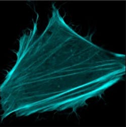
The result of this rational design is a new CFP called mTurquoise2. By combining structural and cellular biology efforts, the researchers showed that mTurquoise2 has a fluorescence efficiency of 93%, enabling study of protein-protein interactions in living cells with unprecedented sensitivity. High sensitivity matters in processes where only a few proteins are involved and signals are weak, and in fast reactions where the time available for accumulating fluorescent light is short.
Thanks to the team's approach, scientists now hope to design improved fluorescent proteins emitting light of different colors for use in other applications, concludes Royant.
The results of the work has been published in Nature Communications; for more information, please visit http://www.nature.com/ncomms/journal/v3/n3/full/ncomms1738.html.
-----
Follow us on Twitter, 'like' us on Facebook, and join our group on LinkedIn
Follow OptoIQ on your iPhone; download the free app here.
Subscribe now to BioOptics World magazine; it's free!


