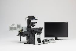
Laser scanning confocal microscope system by Olympus
The FluoView FV1200 laser scanning confocal microscope system from Olympus (Center Valley, PA) for live cell and tissue imaging requires less laser power, resulting in low phototoxicity and photobleaching. Its scanhead features anti-corrosive silver-coated galvo mirrors to increase light throughput and a 748 nm diode laser for near-infrared (NIR) and in-vivo imaging. An optional cooled gallium arsenide phosphide (GaAsP) detector unit delivers 45% quantum efficiency to minimize electrical noise. Multipoint mapping advanced software (MMASW) delivers functional measurements of high-speed calcium fluctuations in groups of cells at speeds up to 101 Hz per field, with sequential position data output up to 50,000 Hz.
-----
High-Sensitivity Olympus FV1200 Biological Laser Scanning Confocal Microscope System Enhances Imaging of Living Specimens
CENTER VALLEY, Pa., October 4, 2012 - Researchers who need more sensitive live cell imaging with high-speed measurement capability will benefit from the Olympus FluoView® FV1200 confocal laser scanning imaging system, designed specifically for optimal live cell and tissue imaging. With its ultra-high sensitivity and innovative fluorescence measurement, it requires less laser power, resulting in lower phototoxicity and photobleaching. Building on Olympus' renowned optical design capability, the FV1200 offers increased laser selection, cooled high-sensitivity detector technology, increased light throughput and faster functional measurements, along with an extraordinary range of high-performance specialty objectives. The system capitalizes on the advantages of the company's recently introduced IX83® automated inverted microscope platform.
Among its many improvements for live cell imaging, the FV1200's scanhead features increased sensitivity via its breakthrough anti-corrosive silver-coated galvo mirrors, which increase light throughput significantly over competitive instruments. The entire system is optimized for sensitivity, with increased excitation and emission transmission efficiencies, particularly in the near-infrared (IR) light range, where much of today's key in vivo and live cell imaging research occurs. The FV1200 also offers a 748nm diode laser for researchers doing near-IR imaging.
Further increasing sensitivity is an optional cooled gallium arsenide phosphide (GaAsP) detector unit that minimizes electrical noise. By using selected GaAsP detectors that deliver 45-percent quantum efficiency, users will experience some of the highest signal-to-noise imaging ratios possible. When the GaAsP unit is used with three conventional confocal detectors, the FV1200 can acquire up to five simultaneous fluorescent channels with its near-IR 748nm laser diode, allowing users to image DAPI, GFP, RPF, Cy5 and Cy7 simultaneously. Combining GaAsP and conventional photomultiplier tubes in one system allows users to select either higher-sensitivity or higher-dynamic-range imaging, providing important flexibility for imaging live cells.
Functional imaging and measurement are also greatly enhanced. The system's multipoint mapping advanced software (MMASW) delivers functional measurements of high-speed calcium fluctuations in groups of cells at speeds up to 101 Hz per field, with sequential position data output up to 50,000 Hz, providing even faster measurements than are possible using raster-scanning resonance scanners. Unlike raster scanning, the pseudo-heuristic point-scanning mode allows users to optimize their scan path as laser light moves from point to point without reducing the field of view by cropping. Each point can be expanded to an array for larger-area stimulation or detection. Competitive scanners that rely on resonance or acousto-optic detectors (AODs) are intrinsically less sensitive due to their very short integration times and the number of photons that can be detected; the Olympus multipoint mode allows greater signal-to-noise performance by retaining high integration time per position in each scan. Precise and accurate multichannel fluorescence measurements are useful for stem cell research, patch clamping and electrophysiology, optogenetic experiments with channelrhodopsin and halorhodopsin, and studies of functional output over cells and cell networks.
The MMASW's mapping feature allows pixel-skipping image generation in a field of fluctuating cells; it also provides automatic region-of-interest identification for further high-speed measurement. Users can make use of the high, physiologically relevant measurement speed while simultaneously stimulating cells in complex multipoint patterns or use the proprietary Olympus tornado scan mode using the optional SIM scanner, a fully functional galvo scanner that operates simultaneously with the main scanner. The SIM scanner can use either single laser-line inputs or the full range of visible lasers using the dual fiber output laser combiner.
Further enhancing its live cell capabilities, the FV1200 provides all the enhanced touch-panel control, upgradeability, increased thermostability and flexibility of the new IX83 microscope system, including its robust implementation of Olympus 'Zero Drift' Compensation (ZDC) technology, which ensures that images always stay in focus. Unlike other focus control systems, ZDC3 allows users to select either the one-shot mode for multi-well applications or constant focus for imaging live specimens over hours or days. The U-shaped IX83 frame design incorporates significant improvements in frame stability and rigidity, providing a solid foundation for live cell experiments. The system is robust, stable and highly expandable.
Additionally, the FV1200 is designed to work with a newly enhanced selection of Olympus live-cell-optimized silicone objectives including 30x, 60x and the brand new 40x high-numerical-aperture silicone immersion objectives; microprobe "stick" objectives; the multiple-award-winning SCALEVIEW objectives, and a super-corrected 60x objective. The 60x super-corrected objective lens is matched precisely to the optics of the FV1200 scanhead, providing tremendous benefits for colocalization analysis.
For more information on the high-sensitivity, high-precision Olympus FV1200 biological laser scanning confocal microscope system, visit www.olympusamerica.com/FV1200 or contact Brendan Brinkman, Olympus America Inc., at 424-298-7402; email [email protected].
About Olympus America Inc., Scientific Equipment Group
Olympus America Scientific Equipment Group provides innovative microscope imaging solutions for researchers, doctors, clinicians and educators. Olympus microscope systems offer unsurpassed optics, superior construction and system versatility to meet the ever-changing needs of microscopists, paving the way for future advances in life science.
About Olympus
Olympus is a precision technology leader, designing and delivering innovative solutions in its core business areas: Medical and Surgical Products, Life Science Imaging Systems, Industrial Measurement and Imaging Instruments and Cameras and Audio Products. Olympus works collaboratively with its customers and affiliates worldwide to leverage R&D investment in precision technology and manufacturing processes across diverse business lines. These include:
-Gastrointestinal endoscopes, accessories, and minimally invasive surgical products;
-Advanced research, clinical and educational microscopes and research and educational digital imaging systems;
-Industrial research, engineering, test, inspection and measuring instruments; and
-Digital cameras and voice recorders.
Olympus serves the healthcare field with integrated product solutions and financial, educational and consulting services that help customers to efficiently, reliably and more easily achieve exceptional results. Olympus develops breakthrough technologies with revolutionary product design and functionality for the consumer and professional photography markets, and also is the leader in gastrointestinal endoscopy and clinical and educational microscopes. For more information, visit www.olympusamerica.com.
-----
Follow us on Twitter
Subscribe now to Laser Focus World magazine; it's free!
