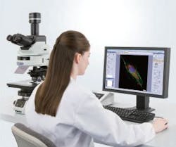
Microscopy software by Olympus
In order to blend together frames, cameras, and optics into a complete microscopy system, Olympus (Hamburg, Germany) has announced the release of a new version of its powerful and flexible cellSens software (now version 1.7). These include the brand new ‘Well Navigator” Solution, which opens up a new world of flexible, multi-well imaging experiments. The software offers High Dynamic Range imaging for perfect exposure of challenging samples, 2D deconvolution for maximizing image clarity and a new database model for streamlining the management of large data repositories. The software now supports additional models of high-sensitivity cameras and is compatible with the Olympus Real Time Controller, which is especially powerful when performing high-end methodologies such as Total Internal Reflection Fluorescence.
-----
PRESS RELEASE
cellSens 1.7 offers multi-well imaging, HDR, 2D deconvolution, real-time control and more
Olympus announces major cellSens software release
Hamburg, 16.07.2012 – Olympus has announced a new release of its powerful and flexible cellSens software (now version 1.7), offering a range of new benefits that allow any user to generate expert results, regardless of experience level. These include the brand new ‘Well Navigator’ Solution, which opens up a new world of flexible, multi-well imaging experiments. cellSens 1.7 also offers High Dynamic Range (HDR) imaging for the perfect exposure of challenging samples, 2D deconvolution for maximising image clarity and a new database model for streamlining the management of large data repositories with many users. The software now supports additional models of high-sensitivity cameras and is compatible with the Olympus Real Time Controller (RTC), which is especially powerful when performing high-end methodologies such as Total Internal Reflection Fluorescence (TIRF).
Olympus cellSens 1.7 is the key element that blends together Olympus frames, cameras and high-quality optics to form a complete microscopy system that is both powerful and effortless to control. A range of easy-to-use tools helps you to get the best out of your life science samples, without the need for extensive expertise. For example, when working with challenging samples that simultaneously contain very light and dark staining, the HDR mode can bring out all the details in the sample with just one click. The new 2D deconvolution tool also improves images by quickly applying a true de-convolution on a single image, without the need for acquiring a Z-stack, for crystal clear images of samples stained with multiple fluorochromes.
This latest release also supports the new optional ‘Well Navigator’ Solution, for the rapid and intuitive set-up of multi-well imaging studies. The system can meet the needs of all users with customizable pre-sets for a wide range of plate formats and panoramic imaging of each well. As focusing is an issue when moving across large multi-well plates this new Solution works seamlessly with the Olympus ZDC focus compensation unit, which can automatically maintain perfect focus without any need for user intervention.
When studying fast dynamic processes a high level of acquisition accuracy is a must for reliable quantitative analysis. For these situations cellSens 1.7 supports a dedicated real-time processor that facilitates advanced imaging via microsecond synchronization of the camera, light source and filter units. The new cellSens 1.7 database has also been redesigned to provide centralised data storage management, optimised for multi-user access. Large groups, or even different institutions, can effortlessly collaborate by securely sharing research data, while maintaining quick and easy access through customizable databases.
There are also a number of user interface enhancements in cellSens 1.7, including the ability to use existing images to set the microscope system to the same acquisition settings, ensuring consistent analysis across many different samples. Furthermore, the new ‘Dark Skin mode’ reduces monitor brightness when performing fluorescent imaging, while maintaining clear coloured icons for intuitive ease-of-use. In combination, the wide range of new benefits offered by this new software release makes cellSens ideal for serving a broad range of life science users, allowing anyone to generate expert results with the minimum of effort.
-----
Follow us on Twitter, 'like' us on Facebook, and join our group on LinkedIn
Follow OptoIQ on your iPhone; download the free app here.
Subscribe now to BioOptics World magazine; it's free!
