Multiphoton microscopy detects skin cancers noninvasively
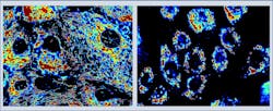
Tufts University (Medford, MA) researchers have developed a noninvasive imaging technique that accurately detects skin cancer without surgical biopsy. Multiphoton microscopy of mitochondria (small organelles that produce energy in cells) accurately identified melanomas and basal cell carcinomas by detecting abnormal clusters of mitochondria in both types of skin cancer.
Related: Multiphoton microscopy records how metastasizing tumor cells move through the body
The researchers, led by Irene Georgakoudi, Ph.D., in the Department of Biomedical Engineering at Tufts University and co-senior author of a paper that describes the work, found that mitochondria behave very differently in healthy vs. cancerous tissue. They used multiphoton microscopy, which takes advantage of the characteristics of a key molecule in mitochondria that is is central to energy production—nicotinamide adenine dinucleotide (NADH). They found that NADH, which naturally fluoresces without injecting any dye or contrast agent into the individuals being screened, can be detected using multiphoton microscopy to provide diagnostically useful information about the organization of the mitochondria in skin cells.
"The system allows us to obtain very high-resolution images of individual cells without having to slice the tissue physically," Georgakoudi explains. "With this technique, we found that in normal cells the mitochondria are spread throughout the cell in a web-like pattern. Conversely, cancerous skin cells show a very different pattern with the mitochondria found in clumps or clusters typically at the center of the cell along the border of the nucleus."
In the research team's study, the technique was tested in 10 patients with skin cancer (melanoma or basal cell carcinoma) and four who did not have skin cancer. The imaging technique results were compared to the traditional biopsy results obtained from each patient. The results demonstrated that the imaging technique correctly identified skin cancer in all 10 cancer patients, and made no false diagnoses in the four individuals without skin cancer.Georgakoudi estimates that this test could be routinely used in doctor's offices within five years, although the $100,000 price tag for the laser used in this microscope could limit the medical facilities that would be able to make such an investment. "Less-expensive lasers are on the horizon," Georgakoudi concludes. "However, this approach would enable a doctor to make a quick diagnosis and begin treatment immediately, which could ultimately lower healthcare costs associated with these very common cancers."
Full details of the work appear in the journal Science Translational Medicine; for more information, please visit http://dx.doi.org/10.1126/scitranslmed.aag2202.

