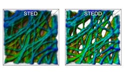
Super-resolution microscopy method suppresses background
Researchers at the Karlsruhe Institute of Technology (KIT; Germany) have developed a super-resolution microscopy method that produces high-resolution images with suppressed background. The new method yields an enhanced image quality that is advantageous when analyzing three-dimensional (3D), densely arranged subcellular structures.
Related: All-fiber illumination brings superior stability to STED
In their work, the researchers refined the stimulated emission depletion (STED) nanoscopy method developed by Stefan W. Hell by modifying image acquisition in a way that suppresses background efficiently. The resulting enhanced image quality is particularly advantageous for quantitative data analysis of 3D molecules and cell structures. The new method, which the researchers call stimulated emission double depletion (STEDD), was developed by Gerd Ulrich Nienhaus of KIT's Institute of Applied Physics (APH) and Institute of Nanotechnology (INT) and his team.
In fluorescence microscopy, the sample to be studied is scanned with a strongly focused light beam to make dye molecules emit fluorescence light. The light quanta are registered pixel by pixel to build up the image. In STED nanoscopy, the excitation beam used for scanning is overlapped by another light beam. Its light intensity is located around the excitation beam and in the center, it is zero. Moreover, the STED beam is shifted towards higher wavelengths.
The STED beam uses the physical effect that was first described by Albert Einstein 100 years ago—namely, stimulated emission—to switch off fluorescent excitation everywhere except in the center where the STED beam has zero intensity. In this way, excitation is confined and a sharper light spot results for scanning. The highly resolved STED image, however, always has a background of low resolution, which is because of incomplete stimulated depletion and fluorescence excitation by the STED beam itself.
The research team has now extended this STED method by another STED beam. The STED2 beam follows the STED beam with a certain time delay and eliminates the useful signal in the center, such that only background excitation remains.
Full details of the work appear in the journal Nature Photonics; for more information, please visit http://dx.doi.org/10.1038/nphoton.2016.279.
