Fluorescence microscopy approach images biological samples with low damage
A team of scientists at the University of St Andrews (St Andrews, Scotland) has developed a fluorescence microscopy method that enables imaging of delicate biological samples with low damage in biomedicine and neuroscience.
Light-sheet fluorescence microscopy allows fast, high-resolution imaging with lower optical damage than other approaches, as it illuminates a sample with a thin sheet of light so other parts of the sample avoid any unnecessary light exposure. The research team has explored how to probe samples in this geometry using much longer wavelengths of illumination.
The team used three units of optical energy (photons) to excite fluorescent labels in the sample. This means they can use much longer excitation wavelengths rather than simply exciting the label directly (one photon) or with two photons. This in turn dramatically reduces light scattering and enhances the penetration of light into the sample.
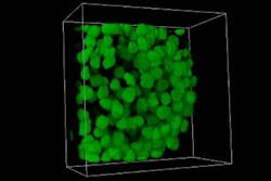
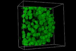
A 3D rendering of a human embryonic kidney is shown.
The researchers imaged spheroids of human embryonic kidney cells using two- and three-photon excitation. At the spheroid’s surface, both imaging modalities performed similarly. However, at the far side of the spheroid, the image quality for the three-photon light-sheet fluorescence microscopy preserved image contrast while the quality of the two-photon image deteriorated considerably. The team further showed how shaping the input mode into a Bessel beam (a pattern with a bright center surrounded by concentric rings) could improve this mode of imaging for the future.
"The use of Bessel beams in three-photon light-sheet fluorescence microscopy will make it possible to image large samples with high resolution, which is crucial for biomedical and neuroscience research," says Adrià Escobet-Montalbán, the lead author of the paper that describes the work.
Full details of the work appear in the journal Optics Letters.

